Review
. 2022 Jun 21;79(7):374.
doi: 10.1007/s00018-022-04408-w.

Inhibition of myostatin and related signaling pathways for the treatment of muscle atrophy in motor neuron diseases

Affiliations
Review

Inhibition of myostatin and related signaling pathways for the treatment of muscle atrophy in motor neuron diseases

Elena Abati et al. Cell Mol Life Sci. .

Abstract

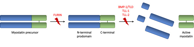
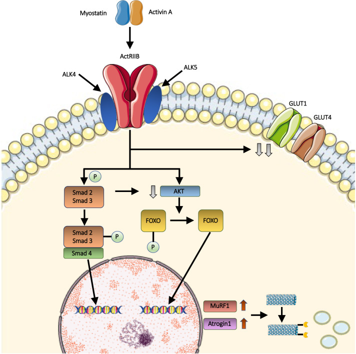
Myostatin is a negative regulator of skeletal muscle growth secreted by skeletal myocytes. In the past years, myostatin inhibition sparked interest among the scientific community for its potential to enhance muscle growth and to reduce, or even prevent, muscle atrophy. These characteristics make it a promising target for the treatment of muscle atrophy in motor neuron diseases, namely, amyotrophic lateral sclerosis (ALS) and spinal muscular atrophy (SMA), which are rare neurological diseases, whereby the degeneration of motor neurons leads to progressive muscle loss and paralysis. These diseases carry a huge burden of morbidity and mortality but, despite this unfavorable scenario, several therapeutic advancements have been made in the past years. Indeed, a number of different curative therapies for SMA have been approved, leading to a revolution in the life expectancy and outcomes of SMA patients. Similarly, tofersen, an antisense oligonucleotide, is now undergoing clinical trial phase for use in ALS patients carrying the SOD1 mutation. However, these therapies are not able to completely halt or reverse progression of muscle damage. Recently, a trial evaluating apitegromab, a myostatin inhibitor, in SMA patients was started, following positive results from preclinical studies. In this context, myostatin inhibition could represent a useful strategy to tackle motor symptoms in these patients. The aim of this review is to describe the myostatin pathway and its role in motor neuron diseases, and to summarize and critically discuss preclinical and clinical studies of myostatin inhibitors in SMA and ALS. Then, we will highlight promises and pitfalls related to the use of myostatin inhibitors in the human setting, to aid the scientific community in the development of future clinical trials.

Keywords: Activin receptors, type II; Monoclonal antibodies; Motor neuron diseases; Muscle atrophy; Myostatin.


Conflict of interest statement

The authors declare no existing conflict of interests.

Figures

Fig. 1
Fig. 1
Schematic representation of myostatin processing. Promyostatin, the inactive precursor of MSTN, is composed by the N-terminal prodomain and the C-terminal dimer. Myostatin activation requires two enzymatic cleavages, operated by the furin proteases and by the BMP/tolloid metalloproteases, respectively, including the bone morphogenetic protein-1/tolloid (BMP-1/TLD), tolloid-like-1 (TLL-1) and tolloid-like-2 (TLL-2). In the latent myostatin, resulted by furin cleavage, the non-covalent binding between the C-terminal and the prodomain prevents myostatin activation. Subsequently, the tolloid cleavage at the aspartate residue 76 allows the release and activation of myostatin
Fig. 2
Fig. 2
Myostatin muscle pathway. The binding of myostatin or, alternatively, activin, to muscle activin receptor type IIB (ActRIIB) results in its dimerization and, subsequently, in the activation of type I activin receptor transmembrane kinases ALK4 or ALK5. Consequently, the Smad2/Smad3 complex is phosphorylated, and the Smad4 component is recruited. The Smad complex enters the nucleus, where it acts as transcriptional activator of downstream genes involved in muscle wasting. Furthermore, the activation of the transmembrane activin receptor leads to the downregulation of AKT, which is involved in FOXO phosphorylation. Dephosphorylated FOXO translocates into the nucleus, and up-regulates the transcription of MuRF1 and Atrogin1. Muscle proteins, which are ubiquitinated by MuRF1 and Atrogin1, are subsequently catabolized by proteasomes, thus resulting in muscle atrophy. Simultaneously, myostatin is involved in glucose homeostasis regulation, likely by reducing the protein levels of glucose transporter 1 (GLUT1) and 4 (GLUT4)

References

    1. D’Amico A, Mercuri E, Tiziano FD, Bertini E. Spinal muscular atrophy. Orphanet J Rare Dis. 2011;6:71. doi: 10.1186/1750-1172-6-71. - DOI - PMC - PubMed
    1. Hardiman O, Al-Chalabi A, Chio A, et al. Amyotrophic lateral sclerosis. Nat Rev Dis Primers. 2017;3:17071. doi: 10.1038/nrdp.2017.71. - DOI - PubMed
    1. Chiò A, Logroscino G, Traynor BJ, et al. Global epidemiology of amyotrophic lateral sclerosis: a systematic review of the published literature. Neuroepidemiology. 2013;41:118–130. doi: 10.1159/000351153. - DOI - PMC - PubMed
    1. Abati E, Bresolin N, Comi G, Corti S. Silence superoxide dismutase 1 (SOD1): a promising therapeutic target for amyotrophic lateral sclerosis (ALS) Expert Opin Ther Targets. 2020;24:295–310. doi: 10.1080/14728222.2020.1738390. - DOI - PubMed
    1. Abati E, Bresolin N, Comi G, Corti S. Advances, challenges, and perspectives in translational stem cell therapy for amyotrophic lateral sclerosis. Mol Neurobiol. 2019;56:6703–6715. doi: 10.1007/s12035-019-1554-x. - DOI - PubMed

MeSH terms