. 2022 Nov;40(11):1156-1165.
doi: 10.1007/s11604-022-01298-7. Epub 2022 Jun 21.

The effect of CT texture-based analysis using machine learning approaches on radiologists' performance in differentiating focal-type autoimmune pancreatitis and pancreatic duct carcinoma

Affiliations

The effect of CT texture-based analysis using machine learning approaches on radiologists' performance in differentiating focal-type autoimmune pancreatitis and pancreatic duct carcinoma

Kenta Anai et al. Jpn J Radiol. 2022 Nov.

Erratum in

Abstract

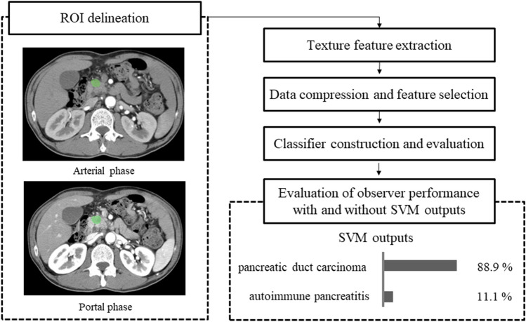
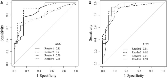
Purpose: To develop a support vector machine (SVM) classifier using CT texture-based analysis in differentiating focal-type autoimmune pancreatitis (AIP) and pancreatic duct carcinoma (PD), and to assess the radiologists' diagnostic performance with or without SVM.

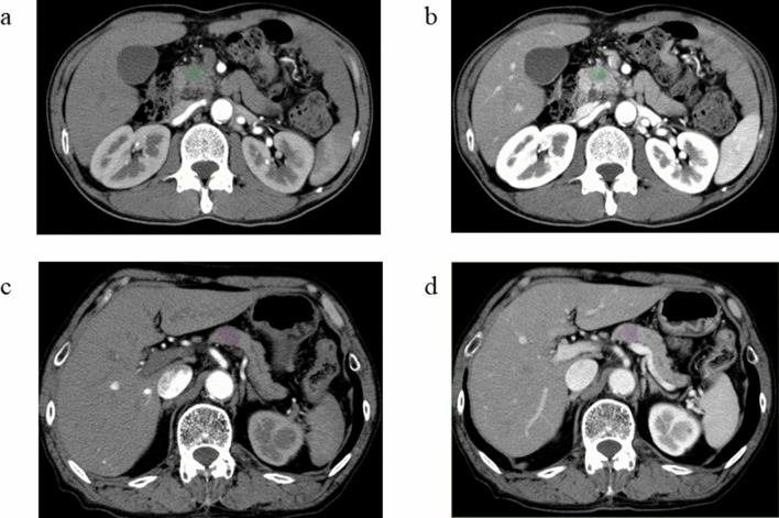
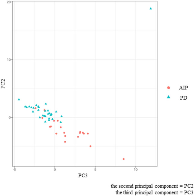
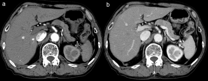
Materials and methods: This retrospective study included 50 patients (20 patients with focal-type AIP and 30 patients with PD) who underwent dynamic contrast-enhanced CT. Sixty-two CT texture-based features were extracted from 2D images of the arterial and portal phase CTs. We conducted data compression and feature selections using principal component analysis (PCA) and produced the SVM classifier. Four readers participated in this observer performance study and the statistical significance of differences with and without the SVM was assessed by receiver operating characteristic (ROC) analysis.

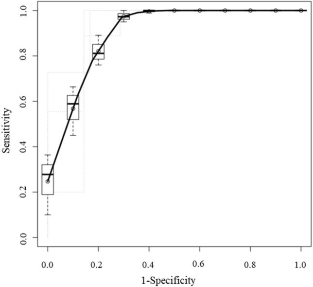
Results: The SVM performance indicated a high performance in differentiating focal-type AIP and PD (AUC = 0.920). The AUC for all 4 readers increased significantly from 0.827 to 0.911 when using the SVM outputs (p = 0.010). The AUC for inexperienced readers increased significantly from 0.781 to 0.905 when using the SVM outputs (p = 0.310). The AUC for experienced readers increased from 0.875 to 0.912 when using the SVM outputs, however, there was no significant difference (p = 0.018).

Conclusion: The use of SVM classifier using CT texture-based features improved the diagnostic performance for differentiating focal-type AIP and PD on CT.

Keywords: Autoimmune pancreatitis; CT; Machine learning; Pancreatic duct carcinoma; Texture analysis.


Conflict of interest statement

We declare that we have no conflicts of interest with any organization or institute.

Figures

Fig. 1
Fig. 1
Overall flowchart explaining the process in this study. First, texture features were extracted from arterial and portal phase CT images using two-dimensional analysis. Then, we conducted data compression and feature selections using principal component (PC) analysis. Subsequently, the support vector machine (SVM) classifier was conducted and the diagnostic accuracy was evaluated. Finally, the effect of SVM on observers’ performance with SVM outputs was also evaluated
Fig. 2
Fig. 2
Arterial phase (a) and portal phase (b) images of a patient with pancreatic duct carcinoma (PD). Arterial phase (c) and portal phase (d) images of a patient with focal-type autoimmune pancreatitis (AIP). A manually defined ROI is drawn in the pancreatic lesion by LIFEx software
Fig. 3
Fig. 3
Scatterplot of the two principal components (PCs). Each dot represents an autoimmune pancreatitis (AIP) (red circle) or a pancreatic duct carcinoma (PD) (blue triangle). Dots were clearly separated between the AIP and the PD groups
Fig. 4
Fig. 4
The receiver operating characteristic curve (ROC) for differentiating focal-type autoimmune pancreatitis (AIP) and pancreatic duct carcinoma (PD) of the support vector machine (SVM) classifier. The areas under the curve (AUC) were 0.920
Fig. 5
Fig. 5
a Receiver operating characteristic curves (ROCs) for differentiating focal-type autoimmune pancreatitis (AIP) and pancreatic duct carcinoma (PD) of all the readers with support vector machine (SVM) outputs. The areas under the curves (AUCs) of the 4 readers were 0.85, 0.90, 0.79, and 0.78, respectively. b ROCs for differentiating focal-type AIP and PD of all the readers with SVM outputs. The AUCs of the 4 readers were 0.91, 0.92, 0.91, and 0.90, respectively
Fig. 6
Fig. 6
Arterial phase (a) and portal phase (b) images of the patient with focal-type autoimmune pancreatitis (AIP). The pancreatic lesion had an absence of atrophic changes in the body and the tail of the pancreas and an absence of significant upstream main pancreatic duct dilatation. The support vector machine (SVM) classified it as a pancreatic duct carcinoma (PD), while all radiologists classified the case correctly as AIP

References

    1. Finkelberg DL, Sahani D, Deshpande V, Brugge WR. Autoimmune pancreatitis. N Engl J Med. 2006;355:2670–2676. doi: 10.1056/NEJMra061200. - DOI - PubMed
    1. Yadav D, Notahara K, Smyrk TC, Clain JE, Pearson RK, Farnell MB, et al. Idiopathic tumefactive chronic pancreatitis: clinical profile, histology, and natural history after resection. Clin Gastroenterol Hepatol. 2003;1:129–135. doi: 10.1053/cgh.2003.50016. - DOI - PubMed
    1. Raina A, Krasinskas AM, Greer JB, Lamb J, Fink E, Moser AJ, et al. Serum immunoglobulin G fraction 4 levels in pancreatic cancer: elevations not associated with autoimmune pancreatitis. Arch Pathol Lab Med. 2008;132:48–53. doi: 10.5858/2008-132-48-SIGFLI. - DOI - PubMed
    1. Kim H-R, Lee C-H, Kim YW, Han SK, Shim Y-S, Yim J-J. Increased CA 19–9 level in patients without malignant disease. Clin Chem Lab Med. 2009;47:750–754. doi: 10.1515/CCLM.2009.152. - DOI - PubMed
    1. La Greca G, Sofia M, Lombardo R, Latteri S, Ricotta A, Puleo S, et al. Adjusting CA19-9 values to predict malignancy in obstructive jaundice: influence of bilirubin and C-reactive protein. World J Gastroenterol. 2012;18:4150–4155. doi: 10.3748/wjg.v18.i31.4150. - DOI - PMC - PubMed

MeSH terms