Clinical Trial
. 2022 Aug 1;132(15):e157726.
doi: 10.1172/JCI157726.

Phase I study of adjuvant immunotherapy with autologous tumor-infiltrating lymphocytes in locally advanced cervical cancer

Affiliations
Clinical Trial

Phase I study of adjuvant immunotherapy with autologous tumor-infiltrating lymphocytes in locally advanced cervical cancer

He Huang et al. J Clin Invest. .

Abstract

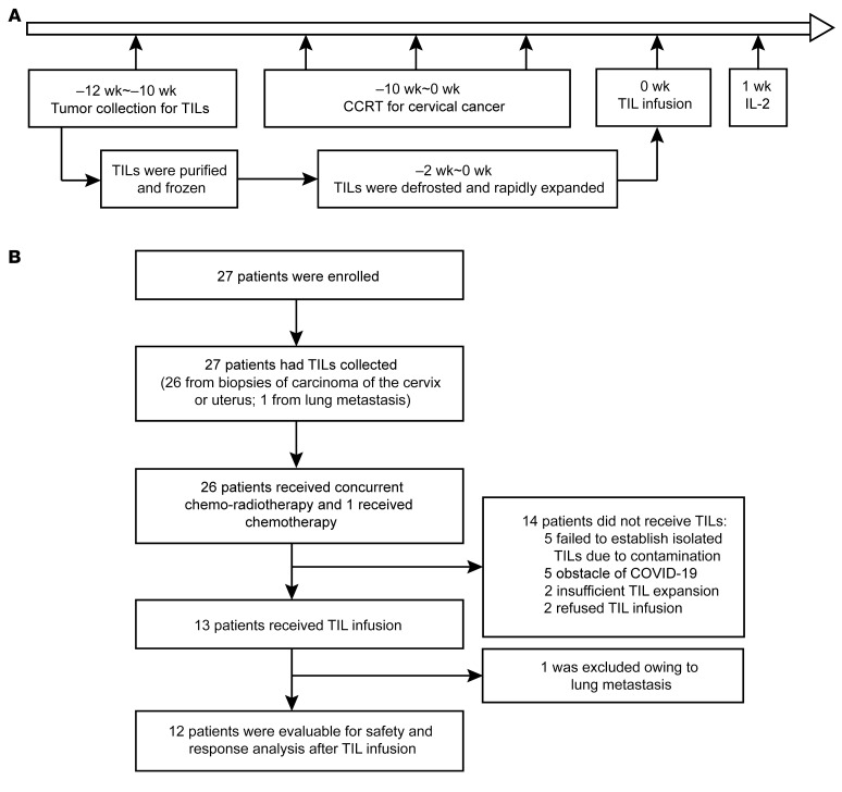
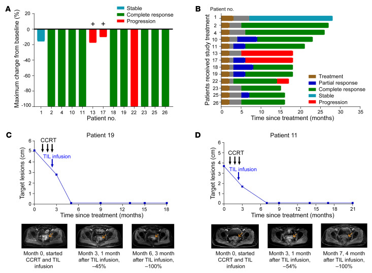
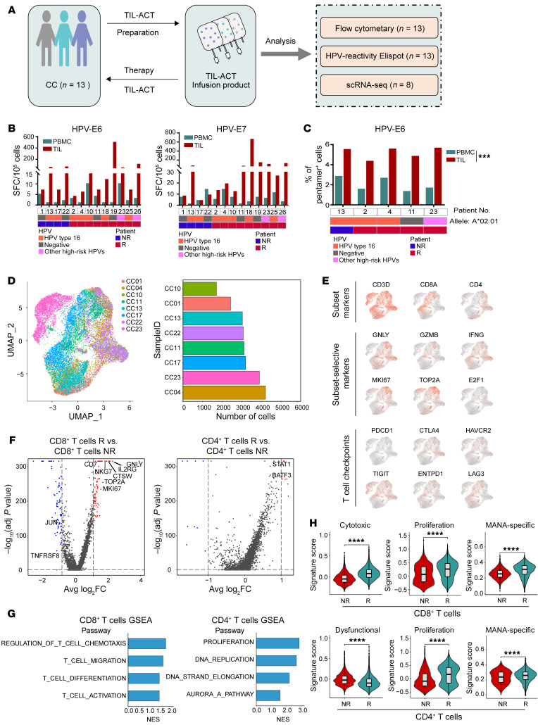
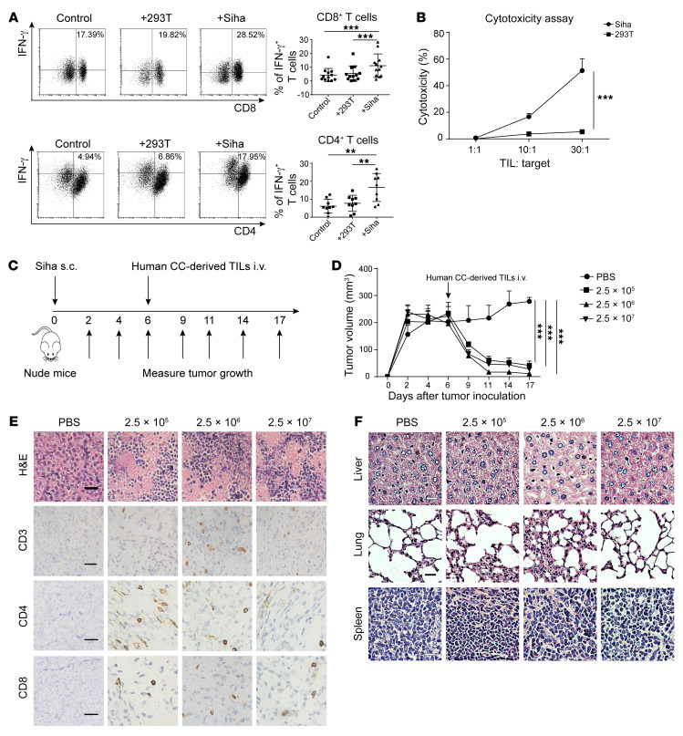
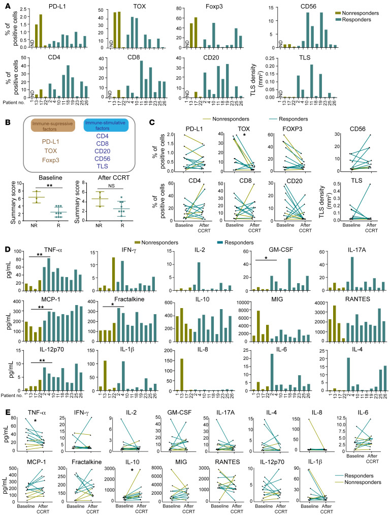
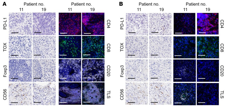
BACKGROUNDAdoptive cell therapy (ACT) with tumor-infiltrating lymphocytes (TILs) has achieved remarkable clinical efficacy in metastatic cancers such as melanoma and cervical cancer (CC). Here, we explored the safety, feasibility, and preliminary tumor response and performed translational investigations of adjuvant immunotherapy using infusion of autogenous TILs (auto-TILs) following concurrent chemoradiotherapy (CCRT) in patients with CC who had locally advanced disease.METHODSTwenty-seven patients with CC with stage III-IV disease were recruited in this single-center, phase I study. TILs were isolated from lesions in the uterine cervix and generated under good manufacturing practice (GMP) conditions and then infused after CCRT plus i.m. IL-2 injections.RESULTSTILs from 20 of the 27 patients were successfully expanded, with a feasibility of 74.1%. Twelve patients received TILs following CCRT. Adverse events (AEs) were primarily attributable to CCRT. Only 1 (8.3%) patient experienced severe toxicity with a grade 3 hypersensitivity reaction after TIL infusion. No autoimmune AEs, such as pneumonitis, hepatitis, or myocarditis, occurred, and there were no treatment-related mortalities. Nine of 12 patients (75.0%) attained a complete response, with a disease control duration of 9-22 months. Translational investigation showed that the transcriptomic characteristics of the infused TIL products and some immune biomarkers in the tumor microenvironment and serum of patients with CC at baseline were correlated with the clinical response.CONCLUSIONTIL-based ACT following CCRT was safe in an academic center setting, with potentially effective responses in patients with locally advanced CC. "Hot" inflammatory immune environments were beneficial to the clinical efficacy of TIL-based ACT as adjuvant therapy.TRIAL REGISTRATIONClinicalTrials.gov NCT04443296.FUNDINGNational Key R&D Program; Sci-Tech Key Program of the Guangzhou City Science Foundation; the Guangdong Province Sci-Tech International Key Program; the National Natural Science Foundation of China.

Keywords: Adaptive immunity; Cancer; Clinical Trials; Oncology.


Figures

Figure 1
Figure 1. Schematic representing the study design and patient disposition.
(A) Clinical trial schema. The week count is relative to TIL infusion. (B) Patient flow chart. Of the 27 patients enrolled, 13 patients received a TIL infusion after CCRT (n = 12 patients) or chemotherapy (n = 1 patient) and were evaluated for safety and tumor response. CCRT, concurrent chemoradiotherapy, radical radiotherapy for CC with concurrent cisplatin 30~40 mg/m2 weekly during EBRT.
Figure 2
Figure 2. Clinical evaluation of patients for CCRT and auto-TIL treatment.
(A) Waterfall plot of the maximum change in the sum of the target lesion (primary tumor lesion of the uterine cervix) compared with baseline measurements in 13 patients. “+” indicates distant metastasis. Patients 13 and 17 presented with distant lung and bone metastases. Patient 22 had pelvic recurrence after a 9-month CR. (B) Swimmer plots of the change in the sum of the target lesions from the treatment in 13 patients. Each bar represents 1 patient in this study. (C and D) MRI scans obtained at baseline and after CCRT and TIL infusion for CC patients 11 and 19.
Figure 3
Figure 3. Correlations of characteristics of infused TIL products and clinical response.
(A) Schematic illustration of biomarkers and functional identification of TIL products in this study. (B) Frequency of T cell reactivity against HPV E6 (left) and E7 (right) antigens in peripheral blood and TILs (n = 13). (C) Frequency of HPV E6 antigen–specific T cells in peripheral blood and in TILs from patients with HLA-A2+ CC (n = 5), by Wilcoxon test. (D) UMAP plot showing cells from 8 patients with CC. Bar graph shows the number of cells for each indicated patient (n = 8). (E) Expression and distribution of canonical T cell marker genes (CD3D, CD8A, and CD4) and genes related to cytotoxicity and proliferation among these cell subsets. (F) Volcano plots showing DEGs in CD8+ T cells (left) and CD4+ T cells (right) in responders versus nonresponders. Representative genes are labeled. adj, adjusted; avg, average; FC, fold change. (G) GSEA shows the pathway activities in CD8+ T cells (left) and CD4+ T cells (right) between responders and nonresponders. NES, normalized enrichment core. (H) Violin plots show the key signature scores of CD8+ T cells (top) and CD4+ T cells (bottom) (responders vs. nonresponders). ***P < 0.001 and ****P < 0.0001, by Mann-Whitney U test. R, responders; NR, nonresponders.
Figure 4
Figure 4. Specific cytotoxic effects and antitumor effects of TILs in vitro and in vivo.
(A) Representative flow cytometric plots (left) and summary graphs (right) showing the frequencies of IFN-γ–producing T cells among CD4+ (n = 9) and CD8+ (n = 12) TILs cocultured with SiHa and 293T cells. (B) LDH cytotoxicity assay showing the specific killing effect of TILs (n = 3). (C) Experimental scheme for monitoring tumor growth and TIL therapy. (D) Time course of tumor growth in different groups adoptively transferred with different doses of human TILs isolated from patients with CC . n = 5. (E) Representative images of H&E staining of transplanted tumor tissue and representative IHC images of staining for anti–human CD3, CD4, and CD8 in the TME. (F) Representative images of H&E staining of liver, lung, and splenic tissue from nude mice in each experimental group. Scale bars: 100 μm. Data are shown as the mean ± SEM. **P < 0.01 and ***P < 0.001, by Mann-Whitney U test (A, B, and D).
Figure 5
Figure 5. Linkage of baseline biomarkers and dynamic changes in biomarkers after CCRT to clinical response.
(A) Percentage of positive cells with indicative biomarkers, including PD-L1, TOX, Foxp3, CD4, CD8, CD56, CD20, and TLSs in 12 tumor specimens from patients with CC at baseline (n = 9 responders and n = 3 nonresponders). (B) Immune factors (top) in the TME were divided into immune-suppressive factors (PD-L1, TOX, Foxp3) and immune-stimulative factors (CD4, CD8, CD20, CD56, TLS) according to the function of the gene or the indicated cell population (bottom). The combined immune score of PD-L1, TOX, Foxp3, CD4, CD8, CD20, CD56, and TLS at baseline (left) and after CCRT (right) in responders (n = 9) versus nonresponders (n = 3). The calculation of the combined immune score is described in Methods. (C) Changes in indicative biomarkers in CC specimens before and after CCRT (n = 12). (D) Histograms showing the serum levels of cytokines and chemokines, including TNF-α, fractalkine, IL-12p70, MCP-1, IFN-γ, IL-2, IL-1b, IL-17a, IL-4, IL-6, GM-CSF, RANTES, IP-10, IL-8, and MIG, at baseline in responders (n = 9) and nonresponders (n = 4). (E) Changes in indicative serum cytokines and chemokines in patients with CC at baseline versus after CCRT (n = 13). *P < 0.05 and **P < 0.01, by Mann-Whitney U test for nonparametric data. A paired Student’s t test was used to determine significance for all comparisons at baseline and after CCRT.
Figure 6
Figure 6. Immune evaluation for tumor microenvironments at baseline and after CCRT from 2 CC patients with CR.
(A and B) Representative IHC and IF images of samples from patients 11 and 19 showing PD-L1, TOX, Foxp3, CD56, CD4 (red), CD8 (green), and CD20 (white) expression, and multiplex IF staining showing TLSs composed of CD20+, CD4+, and CD8+ cells at baseline (A) and after CCRT treatment (B). Scale bars: 50 μm and 100 μm for IHC and IF images, respectively. DAPI (blue) was used for nuclear staining. Original magnification, ×10.

References

    1. Cohen SS, et al. Obesity and screening for breast, cervical, and colorectal cancer in women: a review. Cancer. 2008;112(9):1892–1904. doi: 10.1002/cncr.23408. - DOI - PubMed
    1. Gadducci A, Cosio S. Neoadjuvant chemotherapy in locally advanced cervical cancer: review of the literature and perspectives of clinical research. Anticancer Res. 2020;40(9):4819–4828. doi: 10.21873/anticanres.14485. - DOI - PubMed
    1. Chemoradiotherapy for Cervical Cancer Meta-Analysis Collaboration Reducing uncertainties about the effects of chemoradiotherapy for cervical cancer: a systematic review and meta-analysis of individual patient data from 18 randomized trials. J Clin Oncol. 2008;26(35):5802–5812. doi: 10.1200/JCO.2008.16.4368. - DOI - PMC - PubMed
    1. Wright JD, et al. Prognostic performance of the 2018 International Federation of Gynecology and Obstetrics cervical cancer staging guidelines. Obstet Gynecol. 2019;134(1):49–57. doi: 10.1097/AOG.0000000000003311. - DOI - PMC - PubMed
    1. Creelan BC, et al. Tumor-infiltrating lymphocyte treatment for anti-PD-1-resistant metastatic lung cancer: a phase 1 trial. Nat Med. 2021;27(8):1410–1418. doi: 10.1038/s41591-021-01462-y. - DOI - PMC - PubMed

Publication types

Associated data