. 2023 May 2;119(3):857-866.
doi: 10.1093/cvr/cvac099.

Elucidation of the genetic causes of bicuspid aortic valve disease

Jan Gehlen  1   2 Anja Stundl  3   4   5 Radoslaw Debiec  6   7   8 Federica Fontana  9 Markus Krane  5   10   11 Dinara Sharipova  9 Christopher P Nelson  6   7 Baravan Al-Kassou  3 Ann-Sophie Giel  2 Jan-Malte Sinning  3 Christopher M H Bruenger  2 Carolin F Zelck  2 Laura L Koebbe  2 Peter S Braund  6   7 Thomas R Webb  6   7 Simon Hetherington  12 Stephan Ensminger  13   14 Buntaro Fujita  13   14 Salah A Mohamed  13   14 Malakh Shrestha  15 Heike Krueger  15 Matthias Siepe  16 Fabian Alexander Kari  16 Peter Nordbeck  17 Larissa Buravezky  17 Malte Kelm  18 Verena Veulemans  18 Matti Adam  19 Stephan Baldus  19 Karl-Ludwig Laugwitz  4   5 Yannick Haas  4 Matthias Karck  20 Uwe Mehlhorn  21 Lars Oliver Conzelmann  21 Ingo Breitenbach  22 Corinna Lebherz  23 Paul Urbanski  24 Won-Keun Kim  25 Joscha Kandels  26 David Ellinghaus  27   28 Ulrike Nowak-Goettl  29 Per Hoffmann  1 Felix Wirth  10 Stefanie Doppler  10 Harald Lahm  10 Martina Dreßen  10 Moritz von Scheidt  5   30 Katharina Knoll  5   30 Thorsten Kessler  5   30 Christian Hengstenberg  31 Heribert Schunkert  5   30 Georg Nickenig  3 Markus M Nöthen  1 Aidan P Bolger  7   8   9 Salim Abdelilah-Seyfried  9   32 Nilesh J Samani  6   7 Jeanette Erdmann  14   33 Teresa Trenkwalder  5   30 Johannes Schumacher  1   2
Affiliations

Elucidation of the genetic causes of bicuspid aortic valve disease

Jan Gehlen et al. Cardiovasc Res. .

Abstract

Aims: The present study aims to characterize the genetic risk architecture of bicuspid aortic valve (BAV) disease, the most common congenital heart defect.

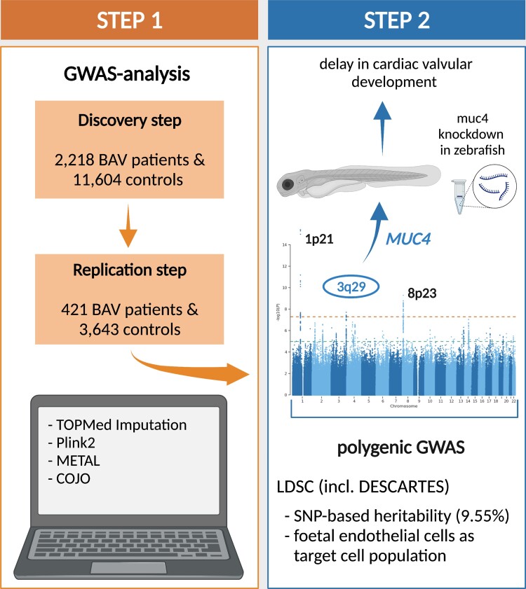
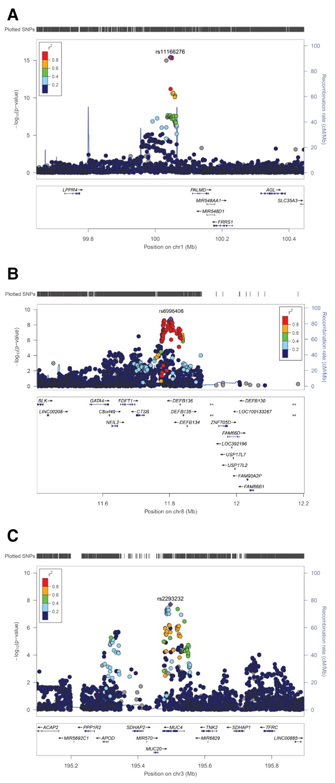
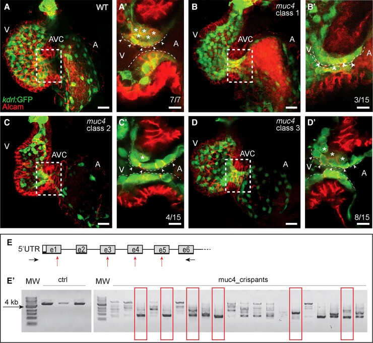
Methods and results: We carried out a genome-wide association study (GWAS) including 2236 BAV patients and 11 604 controls. This led to the identification of a new risk locus for BAV on chromosome 3q29. The single nucleotide polymorphism rs2550262 was genome-wide significant BAV associated (P = 3.49 × 10-08) and was replicated in an independent case-control sample. The risk locus encodes a deleterious missense variant in MUC4 (p.Ala4821Ser), a gene that is involved in epithelial-to-mesenchymal transformation. Mechanistical studies in zebrafish revealed that loss of Muc4 led to a delay in cardiac valvular development suggesting that loss of MUC4 may also play a role in aortic valve malformation. The GWAS also confirmed previously reported BAV risk loci at PALMD (P = 3.97 × 10⁻¹⁶), GATA4 (P = 1.61 × 10-09), and TEX41 (P = 7.68 × 10-04). In addition, the genetic BAV architecture was examined beyond the single-marker level revealing that a substantial fraction of BAV heritability is polygenic and ∼20% of the observed heritability can be explained by our GWAS data. Furthermore, we used the largest human single-cell atlas for foetal gene expression and show that the transcriptome profile in endothelial cells is a major source contributing to BAV pathology.

Conclusion: Our study provides a deeper understanding of the genetic risk architecture of BAV formation on the single marker and polygenic level.

Keywords: Bicuspid aortic valve; Foetal heart transcriptome; GWAS; SNP-based heritability; Zebrafish.


Conflict of interest statement

Conflict of interest: None declared.

Figures

Graphical Abstract
Graphical Abstract
Figure 1.
Figure 1.
Regional association plots of BAV risk loci on chromosome 1p21 near PALMD (A), 8p23 near GATA4 (B) and 3q29 within MUC4 (C). At each locus, associations [−log10(P-values)] are shown for SNPs flanking 400 kb on either side of the lead associated SNP. The lead variant is indicated by the corresponding rs-number. Other markers at each locus are displayed by different colours, which indicate different levels of LD (r2) to the lead SNP. Furthermore, all annotated genes within each region are shown with arrows indicating their transcription direction.
Figure 2.
Figure 2.
Zebrafish muc4 knockout using CRISPR/Cas9. (A–D) Maximum intensity projections from confocal z-stack images of hearts at 58 hpf. (A’–D’) Single confocal z-section images of the AVC region. Endocardial cells in luminal positions (arrowheads) at the AVC are marked by kdrl:GFP and Alcam. Endocardial cells that have already invaded the extracellular matrix and are in abluminal positions are marked with asterisks. (A’) While in WT control embryos many endocardial cells are in abluminal positions, some of the 15 muc4 crispants with a complete truncation of the genomic locus exhibit a delayed ingression of endocardial cells into the cardiac jelly. The severity of the phenotypes varied according to the following classes: (B’) Class 1 embryos (3 of 15) lack an endocardial cell ingression by that stage; (C’) Class 2 embryos (4 of 15), have only a single endocardial cell ingressing into the cardiac jelly at that stage; (D’) in muc4 crispants of class 3 (8 of 15), a few endocardial cells have ingressed into the cardiac jelly. V, ventricle; A, atrium; AVC, atrioventricular canal. Scale bars are 20 µm (A–D) and 10 µm (A’–D’). (E) Schematic representation of the zebrafish muc4 locus and gRNA binding sites (red arrows). Black arrows indicate the positions of primers used to assess the efficacy of CRISPS-induced knockout by PCR. (E’) Single embryo PCR products from control embryos (ctrl) and embryos injected with a mixture of four gRNAs and Cas9 protein (muc4_crispants). Red boxes are indicating samples that lack any WT genomic amplification bands. Among 58 injected embryos that were genotyped, 15 had a large truncation of the genomic region and did not contain any WT band. Only these crispants were used for phenotypic characterization. MW, molecular weight.

References

    1. Siu SC, Silversides CK. Bicuspid aortic valve disease. J Am Coll Cardiol 2010;55:2789–2800. - PubMed
    1. Ward C. Clinical significance of the bicuspid aortic valve. Heart 2000;83:81–85. - PMC - PubMed
    1. Roberts WC, Ko JM. Frequency by decades of unicuspid, bicuspid, and tricuspid aortic valves in adults having isolated aortic valve replacement for aortic stenosis, with or without associated aortic regurgitation. Circulation 2005;111:920–925. - PubMed
    1. Cripe L, Andelfinger G, Martin LJ, Shooner K, Benson DW. Bicuspid aortic valve is heritable. J Am Coll Cardiol 2004;44:138–143. - PubMed
    1. Galian-Gay L, Carro Hevia A, Teixido-Tura G, Rodriguez Palomares J, Gutierrez-Moreno L, Maldonado G, Gonzalez-Alujas MT, Sao-Aviles A, Gallego P, Calvo-Iglesias F, Bermejo J, Robledo-Carmona J, Sanchez V, Saura D, Sevilla T, Burillo-Sanz S, Guala A, Garcia-Dorado D, Evangelista A, BICUSPID investigators . Familial clustering of bicuspid aortic valve and its relationship with aortic dilation in first-degree relatives. Heart 2019;105:603–608. - PubMed

Publication types

MeSH terms