. 2022 Sep 1;206(5):596-607.
doi: 10.1164/rccm.202106-1503OC.

Thromboxane-Prostanoid Receptor Signaling Drives Persistent Fibroblast Activation in Pulmonary Fibrosis

Affiliations

Thromboxane-Prostanoid Receptor Signaling Drives Persistent Fibroblast Activation in Pulmonary Fibrosis

Toshio Suzuki et al. Am J Respir Crit Care Med. .

Abstract

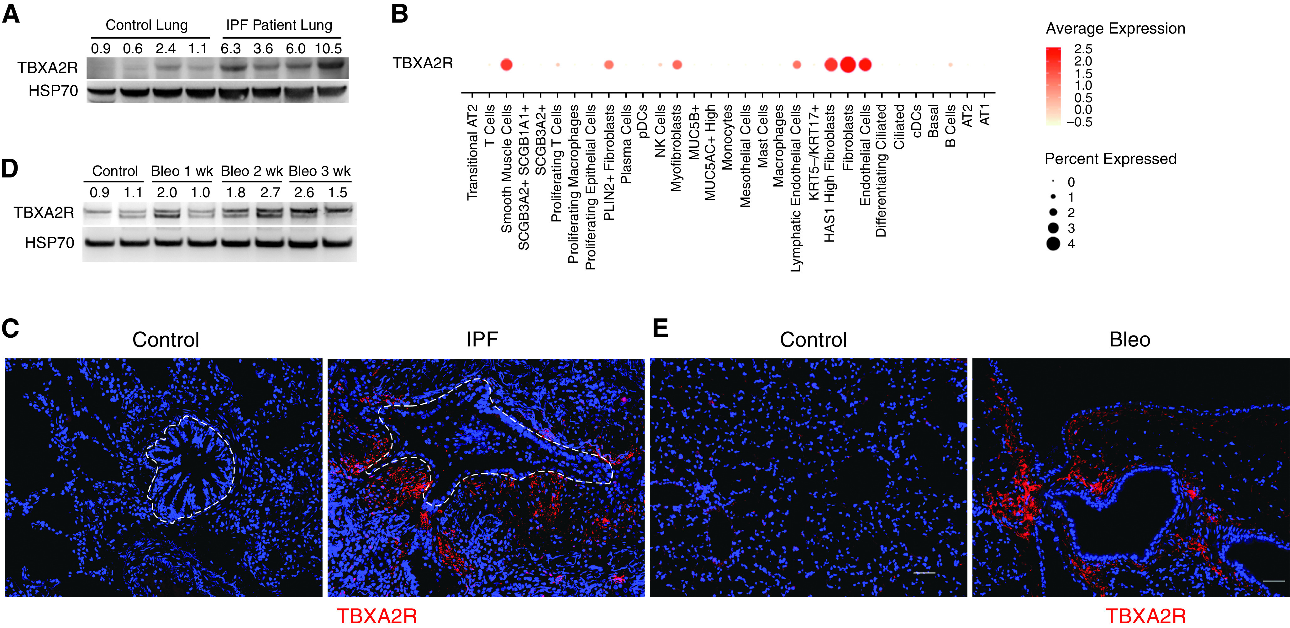
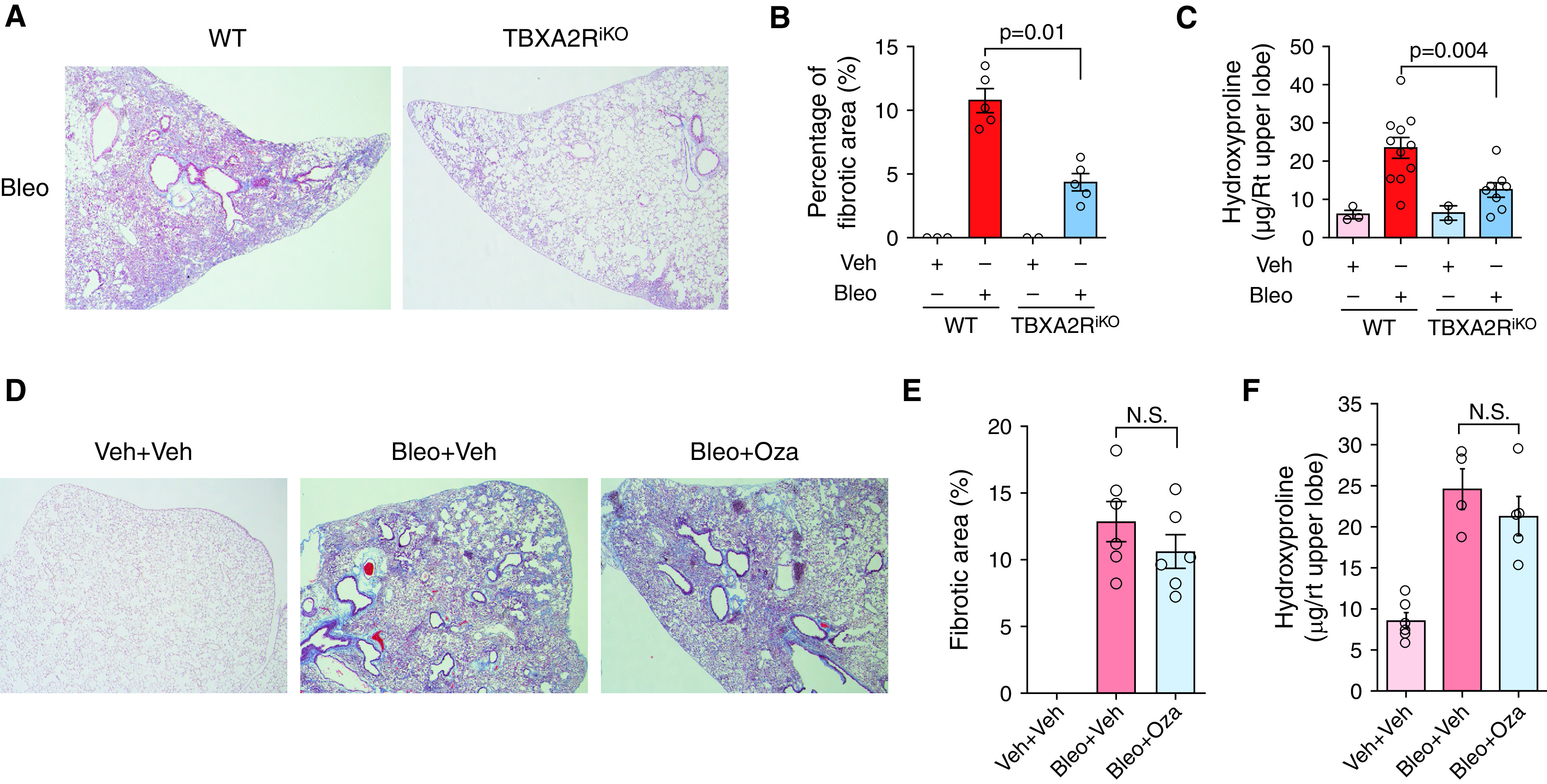
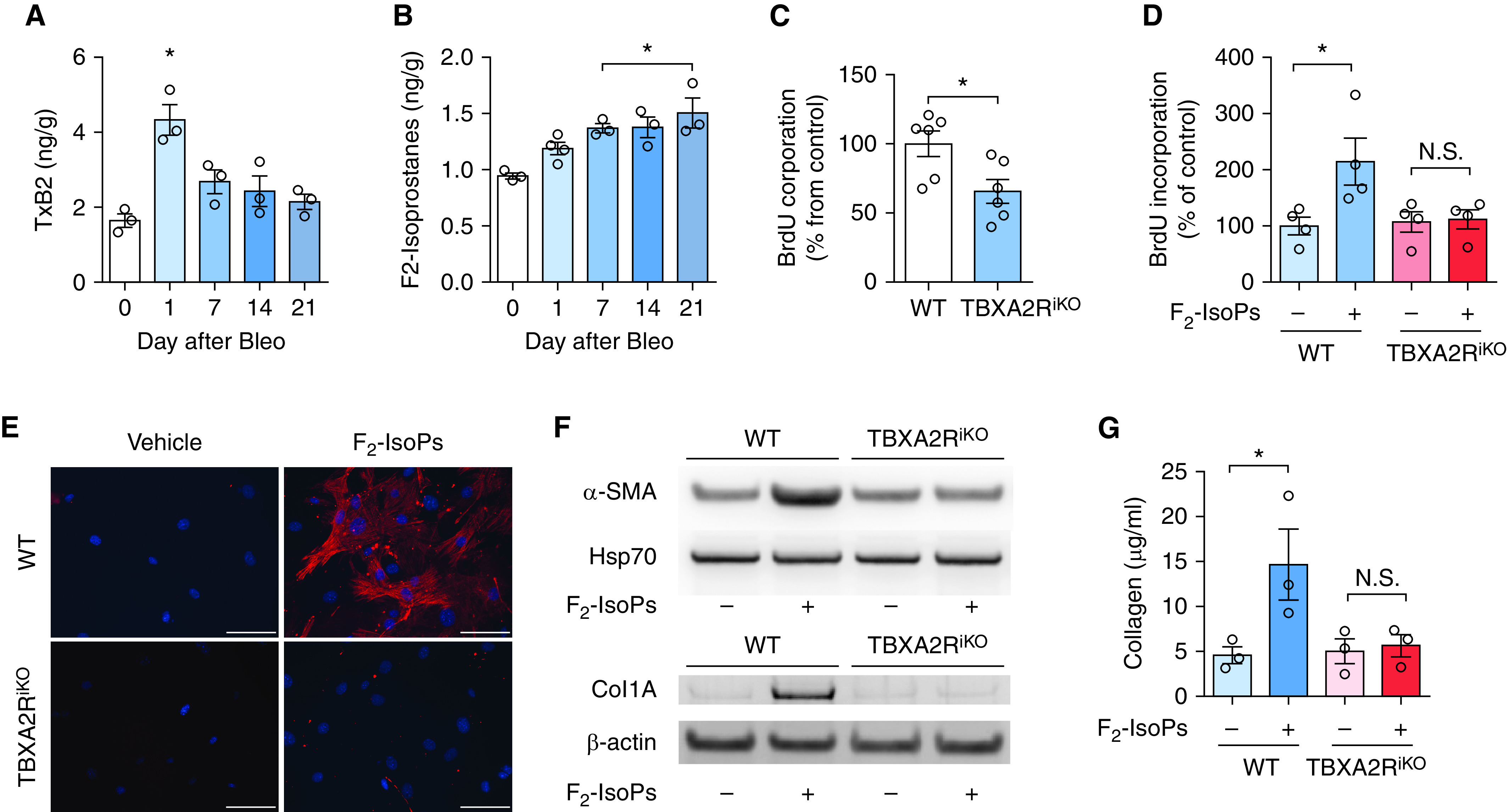
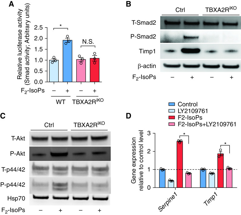
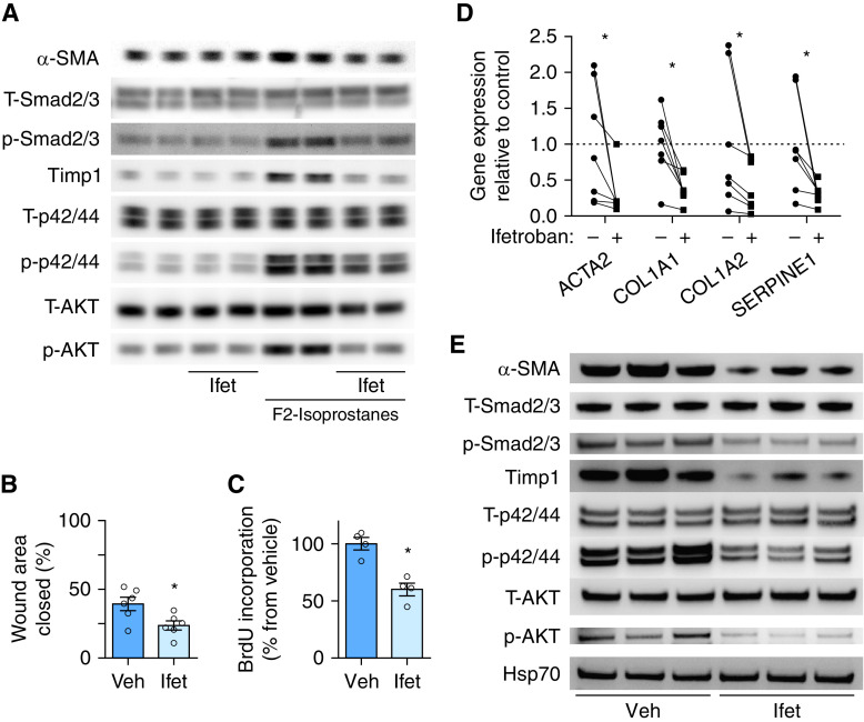
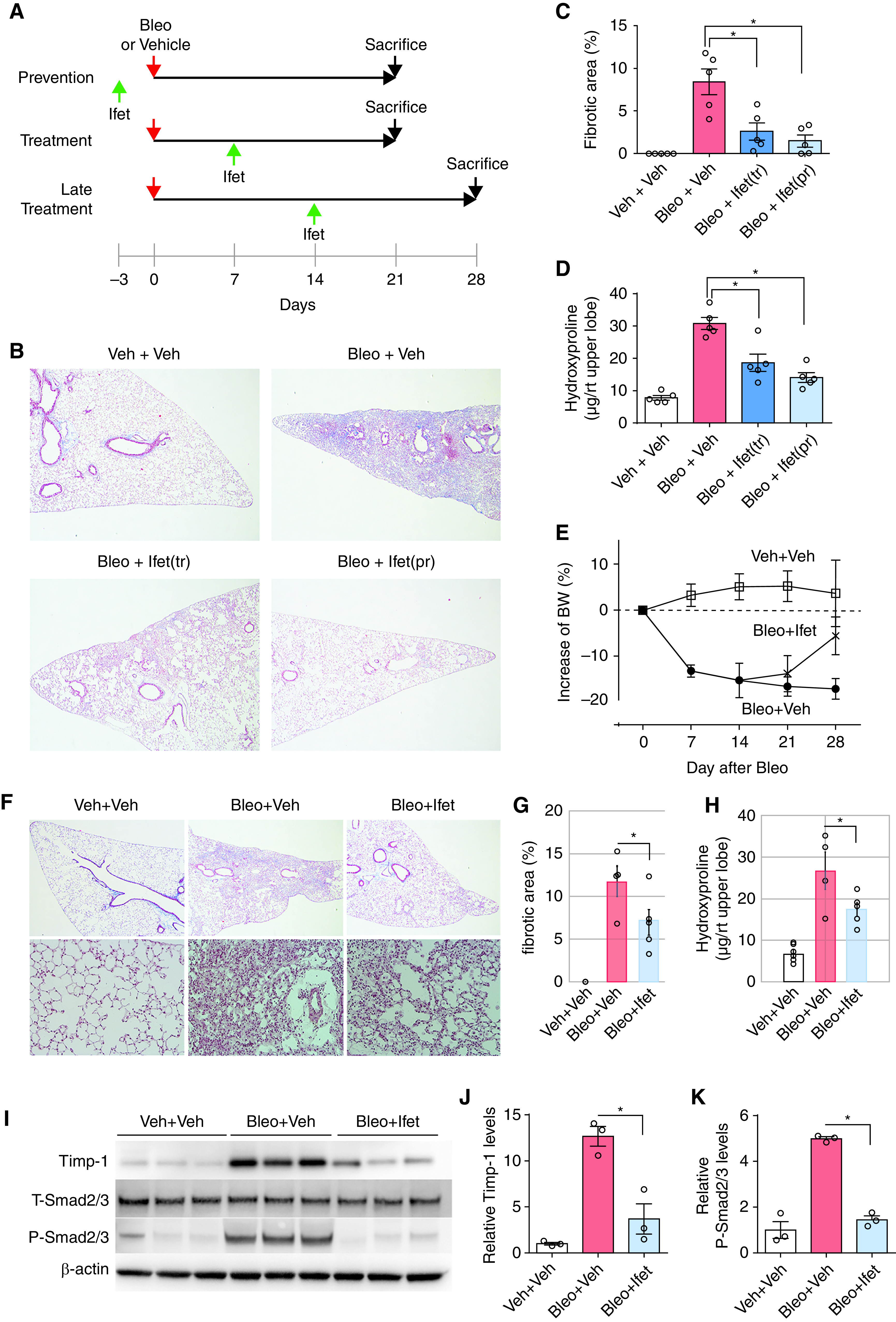
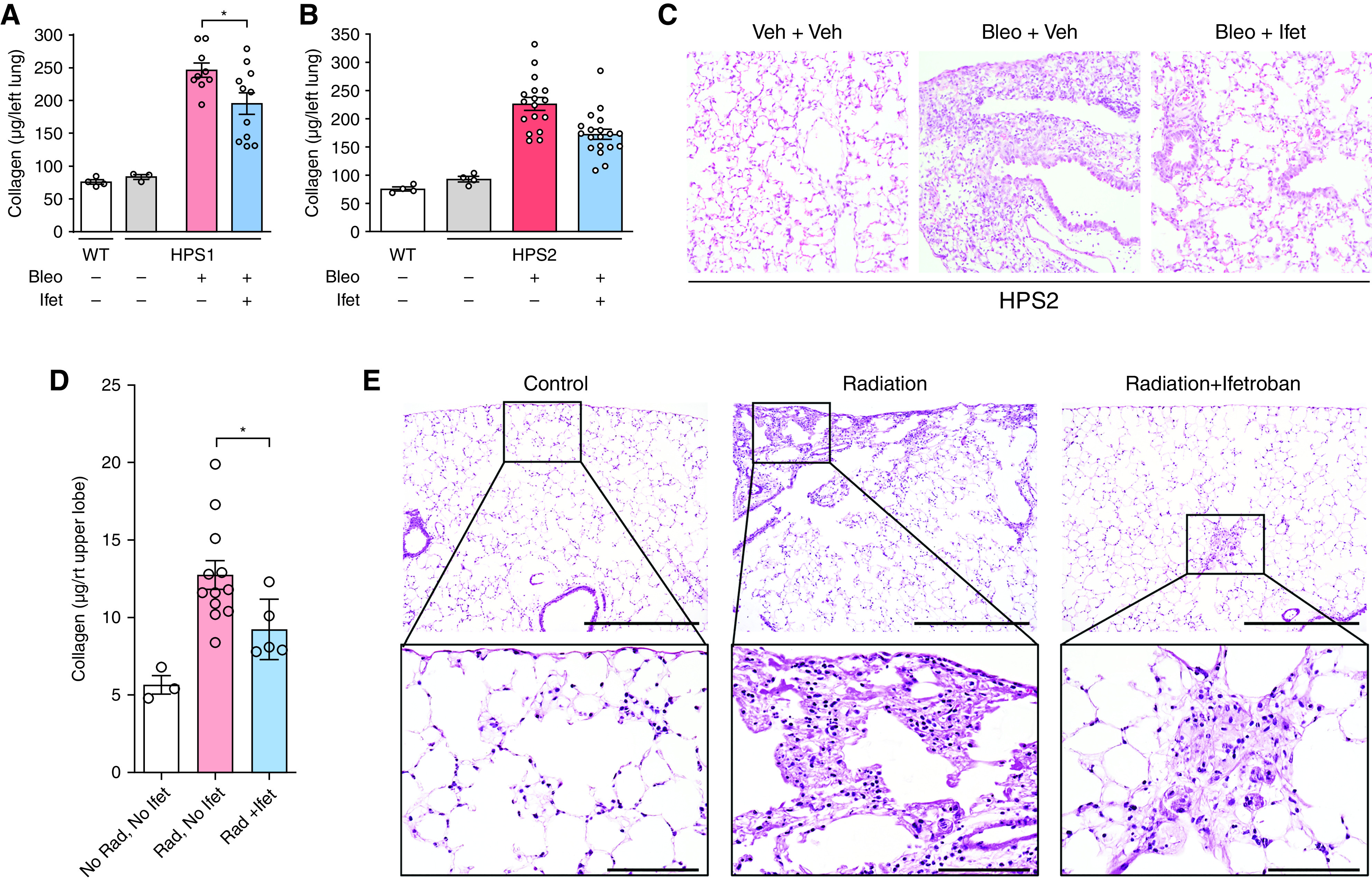
Rationale: Although persistent fibroblast activation is a hallmark of idiopathic pulmonary fibrosis (IPF), mechanisms regulating persistent fibroblast activation in the lungs have not been fully elucidated. Objectives: On the basis of our observation that lung fibroblasts express TBXA2R (thromboxane-prostanoid receptor) during fibrosis, we investigated the role of TBXA2R signaling in fibrotic remodeling. Methods: We identified TBXA2R expression in lungs of patients with IPF and mice and studied primary mouse and human lung fibroblasts to determine the impact of TBXA2R signaling on fibroblast activation. We used TBXA2R-deficient mice and small-molecule inhibitors to investigate TBXA2R signaling in preclinical lung fibrosis models. Measurements and Main Results: TBXA2R expression was upregulated in fibroblasts in the lungs of patients with IPF and in mouse lungs during experimental lung fibrosis. Genetic deletion of TBXA2R, but not inhibition of thromboxane synthase, protected mice from bleomycin-induced lung fibrosis, thereby suggesting that an alternative ligand activates profibrotic TBXA2R signaling. In contrast to thromboxane, F2-isoprostanes, which are nonenzymatic products of arachidonic acid induced by reactive oxygen species, were persistently elevated during fibrosis. F2-isoprostanes induced TBXA2R signaling in fibroblasts and mediated a myofibroblast activation profile due, at least in part, to potentiation of TGF-β (transforming growth factor-β) signaling. In vivo treatment with the TBXA2R antagonist ifetroban reduced profibrotic signaling in the lungs, protected mice from lung fibrosis in three preclinical models (bleomycin, Hermansky-Pudlak mice, and radiation-induced fibrosis), and markedly enhanced fibrotic resolution after bleomycin treatment. Conclusions: TBXA2R links oxidative stress to fibroblast activation during lung fibrosis. TBXA2R antagonists could have utility in treating pulmonary fibrosis.

Keywords: Hermansky-Pudlak syndrome; IPF; oxidative stress; thromboxane–prostanoid receptor; transforming growth factor-β.


Figures

Figure 1.
Figure 1.
TBXA2R (thromboxane–prostanoid receptor) is upregulated in fibroblasts during lung fibrosis. (A) Western blot nondiseased for TBXA2R in lung tissue from patients with idiopathic pulmonary fibrosis (IPF) compared with nondiseased control subjects. (B) Dot plot depicting TBXA2R gene expression in jointly analyzed single-cell RNA sequencing data from explanted lungs of 20 patients with pulmonary fibrosis and 10 nondiseased control subjects (primary data from Reference 6). (C) Representative immunostaining for TBXA2R in IPF and control lung sections. TBXA2R is red, and vimentin is green. (D) Western blot for TBXA2R expression in lungs of mice at baseline and 1–3 weeks after intratracheal bleomycin. (E) Immunostaining for TBXA2R in mouse lung sections at 3 weeks after treatment with vehicle (control) or bleomycin. TBXA2R is red. Scale bar, 50 μm. AT1 = alveolar type 1; AT2 = alveolar type 2; Bleo = bleomycin; cDC = classical dendritic cell; HAS1 = hyaluronan synthase 1; HSP70 = heat shock protein 70; KRT5 = keratin 5; KRT17 = keratin 17; MUC5AC = mucin 5AC, oligomeric mucus/gel-forming; MUC5B = mucin 5B, oligomeric mucus/gel-forming; NK = natural killer; pDC = plasmacytoid dendritic cell; PLIN2 = perilipin 2; SCGB1A1 = secretoglobin family 1A member 1; SCGB3A2 = secretoglobin family 3A member 2.
Figure 2.
Figure 2.
Deleting TBXA2R (thromboxane–prostanoid receptor) protects against experimental lung fibrosis. (A) Representative Masson’s trichrome–stained lung sections from wild-type (WT) and TBXA2RiKO (Rosa26-CreER+TBXA2Rf/f) mice at 21 days after intratracheal bleomycin (Bleo). (B) Morphometric evaluation of lung fibrosis on lung sections. n = 3 for control WT and TBXA2RiKO mice, and n = 5 for the Bleo treatment groups. (C) Hydroxyproline content in the right upper lobe. n = 3 for control WT mice, n = 2 for control TBXA2RiKO mice, n = 11 for the Bleo-treated WT mice, and n = 8 for the Bleo-treated TBXA2RiKO mice. Statistical comparison was performed using the Wilcoxon rank sum test (also P < 0.05 by two-way ANOVA with Tukey’s honestly significant difference test). (D) Representative Masson’s trichrome–stained lung sections from WT mice at 21 days after treatment with intratracheal Bleo or vehicle (Veh), with or without thromboxane synthesis inhibitor (Oza) beginning on Day 7. (E) Morphometric evaluation of lung fibrosis on lung sections. n = 6 for each group. (F) Hydroxyproline content in right upper lobe. n = 6 for each group. Error bars in all panels denote SEM. N.S. = not significant by Wilcoxon rank sum test; Oza = ozagrel; Rt = right.
Figure 3.
Figure 3.
F2-isoprostanes (F2-IsoPs) activate TBXA2R signaling in fibrosis. (A) TxB2 was measured in the lungs of mice at baseline and 1–3 weeks after bleomycin (Bleo). n = 3 per group. *P < 0.05 compared with baseline. (B) F2-IsoPs measured in the lungs of mice at baseline and 1–3 weeks after intratracheal Bleo. n = 3 or 4 per group. *P < 0.05 compared with baseline. (C) Primary lung fibroblasts from Rosa-creER/Tbxa2rf/f mice were treated ex vivo with 4OH-tamoxifen to obtain TBXA2R-deleted fibroblasts (TBXA2RiKO) and BrdU incorporation was measured at 48 hours under full-serum (10%) conditions. n = 6 mice per group. *P < 0.05 compared with wild-type (WT) fibroblasts. (D) F2-IsoP (100 nM) stimulation of WT and Tbxa2riKO fibroblasts under reduced serum conditions (2.5%) measured at 48 hours. n = 4 per group. *P < 0.05. (E) Immunocytochemistry evaluating for α-SMA (α-smooth muscle actin) expression 48 hours after F2-IsoP treatment. α-SMA, red; DAPI, blue. Scale bars, 40× image with 100 μm. (F) Western blots for α-SMA and collagen 1 expression at 48 hours after F2-IsoP treatment. (G) Densitometry for collagen 1. n = 3 lungs per condition. All comparisons were significant both by nonparametric (Wilcoxon rank sum) and parametric (ANOVA with post hoc test) measures. Error bars denote SEM. BrdU = bromodeoxyuridine; Hsp70 = heat shock protein 70; N.S. = not significant; TxB2 = thromboxane B2; TBXA2R = thromboxane–prostanoid receptor.
Figure 4.
Figure 4.
F2-isoprostane (F2-IsoP) induction of the TGF-β pathway requires TBXA2R. (A) Luciferase assay was performed to quantify Smad2/3 transcriptional activity after transfection with Smad2/3-binding element–driven luciferase reporter construct using wild-type (WT) and Tbxa2riKO (Rosa26-CreER+TBXA2Rf/f) lung fibroblasts at 8 hours after F2-IsoP or vehicle treatment. n = 3 for each group. *P < 0.05. (B) Western blot for T-Smad2, P-Smad2, and Timp1 (tissue inhibitor of metalloproteinase-1) using WT and Tbxa2riKO lung fibroblasts at 24 hours after F2-IsoP or vehicle treatment. (C) Western blot for T-Akt, P-Akt, T-p44/42, and P-p44/42 using WT and Tbxa2riKO lung fibroblasts at 24 hours after F2-IsoP or vehicle treatment. Western blots were performed in triplicate samples and repeated at least twice. (D) WT mouse lung fibroblasts were treated with or without F2-IsoPs (100 nM) and LY2109761, an inhibitor of TGF-β receptor type 1/type II kinases, for 24 hours. Quantitative PCR analysis for Serpine1 and Timp1 gene expression was performed. n = 3 for each group. *P < 0.05. Significance was determined using the Wilcoxon rank sum test. Error bars denote SEM. Ctrl = control; Hsp70 = heat shock protein 70; N.S. = not significant; P-Akt = phospho-Akt; P-p44/42 = phospho-p44/42; P-Smad2 = phospho-Smad2; SERPINE1 = serpin family E member 1; T-Akt = total Akt; TBXA2R = thromboxane–prostanoid receptor; TGF-β = transforming growth factor-β; T-p44/42 = total p44/42; T-Smad2 = total Smad2.
Figure 5.
Figure 5.
TBXA2R antagonism reduces proliferation, migration, and activation of fibroblasts from lungs of mice and patients with IPF. (A) Lung fibroblasts were isolated from lungs of mice and treated with F2-isoprostanes, ifetroban (Ifet), or both. Western blots are shown for evaluation of α-SMA, total Smad2/3 (T-Smad2/3), phospho-Smad2/3 (P-Smad2/3), Timp1 (tissue inhibitor of metalloproteinase-1), total p44/42 (T-p44/42), phospho-p44/42 (P-p44/42), total Akt (T-AKT), and phospho-Akt (P-AKT). (B–E) Lung fibroblasts were isolated from explanted lungs of six patients with IPF and treated with Ifet (300 nM) or vehicle. (B) Scratch wound closure assay measured as wound area closed at 24 hours. *P < 0.05 by Wilcoxon signed rank test. Error bars denote SEM. (C) Cell proliferation measured by BrdU incorporation. *P < 0.05 by Wilcoxon signed rank test. Error bars denote SEM. (D) Quantitative PCR for evaluation of profibrotic gene expression. Lines connect cells from the same patient with and without Ifet. Average of vehicle controls was set to 1. n = 6 per group. *P < 0.05 compared with vehicle-treated group by paired t test (each treated culture was paired to the same culture with no treatment). (E) Western blot for evaluation of T-Smad2/3, P-Smad2/3, Timp1, T-p44/42, P-p44/42, T-AKT, and P-AKT. ACTA2 = actin alpha 2, smooth muscle; α-SMA = α-smooth muscle actin; BrdU = bromodeoxyuridine; COL1A1 = collagen type I alpha 1 chain; COL1A2 = collagen type I alpha 2 chain; Hsp70 = heat shock protein 70; IPF = idiopathic pulmonary fibrosis; SERPINE1 = serpin family E member 1; TBXA2R = thromboxane–prostanoid receptor; Veh = vehicle.
Figure 6.
Figure 6.
TBXA2R signaling regulates bleomycin (Bleo)–induced fibrosis, even when started 7 and 14 days after injury. (A) Experimental schema showing the timing of ifetroban (Ifet) start and date of sacrifice for panels. The top two lines correspond to BD in this figure and the bottom line to EK. (B–D) Wild-type mice were treated with intratracheal Bleo (0.04 U) or vehicle (Veh), with or without Ifet (25 mg/kg/day). Ifet treatment (Ifet[tr]) was delivered from Day 7 to Day 21 after Bleo. Ifet prevention (Ifet[pr]) was started 3 days before Bleo treatment and continued to Day 21. (B) Representative Masson’s trichrome–stained lung sections. (C) Morphometric evaluation of lung fibrosis using these lung sections. (D) Hydroxyproline content in right upper lobe. *P < 0.05 by Wilcoxon signed rank test. (E) Change in weight after Bleo treatment. Data are presented as mean ± SEM. Significance was determined using ANOVA with repeated measures. (F) Representative Masson’s trichrome–stained lung sections from wild-type mice at 28 days after treatment with intratracheal Bleo or Veh with or without Ifet treatment begun at Day 14. (G) Morphometric evaluation of lung fibrosis on lung sections. Data are presented as mean ± SEM. (H) Hydroxyproline content quantified from the right upper lobe. Data are presented as mean ± SEM. *P = 0.05 by Wilcoxon signed rank test. (I) Western blot for Timp-1 (tissue inhibitor of metalloproteinase-1), phospho-Smad2/3, and total Smad2/3 in lung tissue. (J) Densitometry for Timp-1. *P < 0.05 by Wilcoxon signed rank test. (K) Densitometry for total and phospho-Smad2/3 (n = 5 per group). *P < 0.05 by Wilcoxon signed rank test. Error bars denote SEM. BW = body weight; P-Smad2/3 = phospho-Smad2/3; rt = right; TBXA2R = thromboxane–prostanoid receptor; T-Smad2/3 = total Smad2/3.
Figure 7.
Figure 7.
TBXA2R antagonism attenuates Hermansky-Pudlak syndrome (HPS)-associated fibrosis and radiation-induced fibrosis. (A and B) HPS1, HPS2, or wild-type control mice were treated with ifetroban (Ifet) or vehicle, followed by intratracheal bleomycin (0.025 U), and lungs were harvested on Day 7. Sircol assay was conducted to quantify collagen content in lungs of HPS1 mice or HPS2 mice. n = 3 or 4 per group unchallenged mice and 9–17 per group HPS mice. (C) Representative histology for HPS2 mice with or without Ifet pretreatment. (D and E) Sircol assay quantifying collagen content (D) and representative histology from lungs of mice exposed to thoracic irradiation (17 Gy) together with Ifet (n = 5) or vehicle (n = 12) and harvested at 4 months after irradiation (E). Top row, scale bars, 4× image with 1000 μm; bottom row, scale bars, 40× with 100 μm. *P < 0.05. For all quantitative figure elements, data are presented as mean ± SEM, with data from each individual mouse overlaid as circles. Statistical analysis was performed using ANOVA, followed by Tukey’s honestly significant difference test for comparisons between individual groups (*P < 0.05). Bleo = bleomycin; Rad = radiation; TBXA2R = thromboxane–prostanoid receptor; Veh = vehicle; WT = wild-type.

Comment in

References

    1. Lederer DJ, Martinez FJ. Idiopathic pulmonary fibrosis. N Engl J Med . 2018;378:1811–1823. - PubMed
    1. Hewlett JC, Kropski JA, Blackwell TS. Idiopathic pulmonary fibrosis: epithelial-mesenchymal interactions and emerging therapeutic targets. Matrix Biol . 2018;71-72:112–127. - PMC - PubMed
    1. Pardo A, Selman M. Idiopathic pulmonary fibrosis: new insights in its pathogenesis. Int J Biochem Cell Biol . 2002;34:1534–1538. - PubMed
    1. Winters NI, Burman A, Kropski JA, Blackwell TS. Epithelial injury and dysfunction in the pathogenesis of idiopathic pulmonary fibrosis. Am J Med Sci . 2019;357:374–378. - PMC - PubMed
    1. Adams TS, Schupp JC, Poli S, Ayaub EA, Neumark N, Ahangari F, et al. Single cell RNA-seq reveals ectopic and aberrant lung-resident cell populations in idiopathic pulmonary fibrosis. Sci Adv . 2020;6:eaba1983. - PMC - PubMed

Publication types