

Analysis of Thrombin-Activated Platelet-Derived Exosome (T-aPDE) Potential for Dental Pulp Regeneration: In-Vitro Study
- PMID: 35728610
- PMCID: PMC9949920
- DOI: 10.1055/s-0042-1744370
Analysis of Thrombin-Activated Platelet-Derived Exosome (T-aPDE) Potential for Dental Pulp Regeneration: In-Vitro Study
Abstract
Objective: This study analyzed the potential of various concentrations of the thrombin-activated platelet-derived exosome (T-aPDE) to regenerate the dental pulp by performing an in-vitro analysis of the cell viability, migration activity, and vascular endothelial growth factor A (VEGF-A) expression of human dental pulp stem cells (hDPSCs).
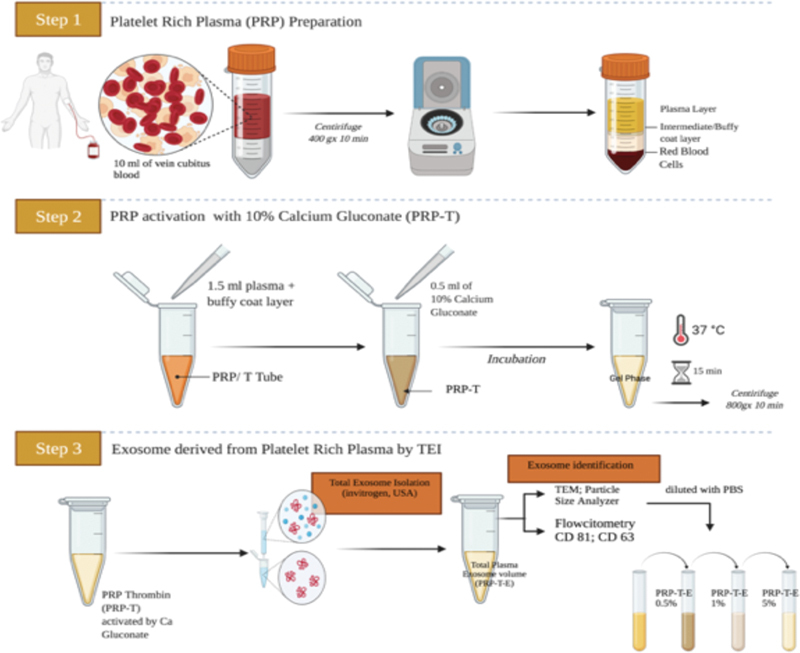
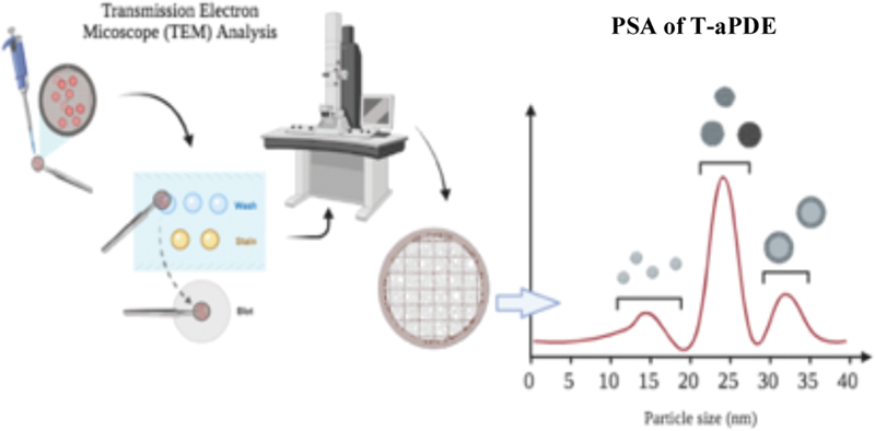
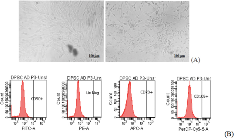
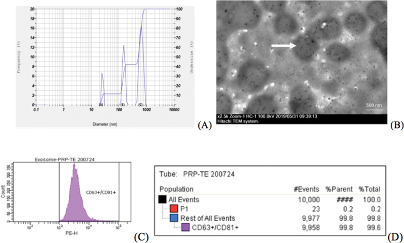
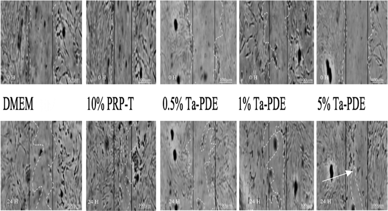
Material and methods: The hDPSCs were collected from nine third molar teeth of nine healthy donors and were isolated and cultured using the explant method. They were harvested between the third and fourth passages and starved, after which they were seeded in the following treatments: Dulbecco's Modified Eagle Medium and 10% platelet-rich plasma-thrombin as the control groups, and 0.5, 1, and 5% T-aPDE as the experimental groups. All groups had three biological triplicates (Triplo) and two number of experiments. The T-aPDE was analyzed using transmission electron microscopy testing, particle size analyzer, and CD63 + and CD81 + specific immune phenotyping flow cytometry tests for plasma exosomes. The cell viability was evaluated using the colorimetric assay of activity cellular enzymes (MTT assay); the migration activity, using scratch assay; and the VEGF-A expression, using enzyme-linked immunosorbent assay.
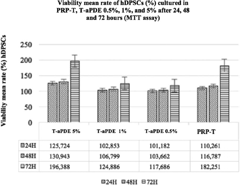
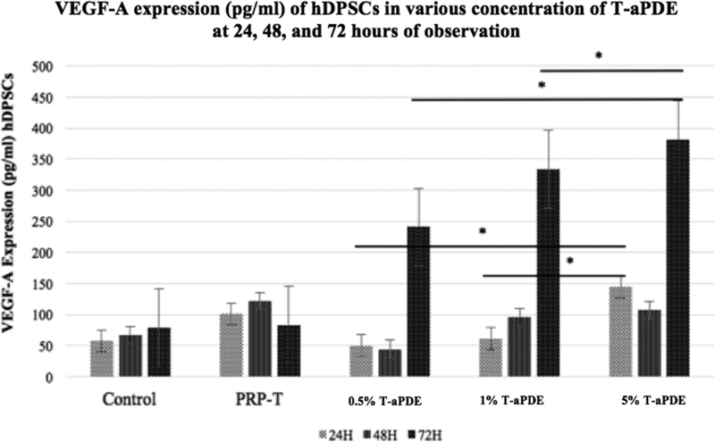
Results: The highest viability absorbance value of hDPSCs after 24, 48, 72 hours of observation was in the 5% T-aPDE group (p<0.05). Whereas, the closest distance result of migratory activation hDPSCs was also in the same group (p<0.05). However the highest VEGF-A expression of hDSPCs was noted in the same group at 72 hours observation (p<0.05).
Statistical analysis: The data were analyzed using one-way analysis of variance and the Kruskal-Wallis test. The statistical power was set at p <0.05 CONCLUSION: The 5% T-aPDE had a higher potential to induce dental pulp regeneration than the other groups.
The Author(s). This is an open access article published by Thieme under the terms of the Creative Commons Attribution License, permitting unrestricted use, distribution, and reproduction so long as the original work is properly cited. (https://creativecommons.org/licenses/by/4.0/).
Conflict of interest statement
None declared.
Figures
References
-
- Žižka R, Šedý J. Paradigm shift from stem cells to cell free regenerative endodontic procedures: a critical review. Stem Cells Dev. 2017;26(03):147–153. - PubMed
-
- Kim S G, Malek M, Sigurdsson A, Lin L M, Kahler B. Regenerative endodontics: a comprehensive review. Int Endod J. 2018;51(12):1367–1388. - PubMed
-
- American Association of Endodontists Scope of endodontics: regenerative endodonticsAccessed November 2, 2020 at:https://www.aae.org/specialty/wp-content/uploads/sites/2/2017/06/scopeof...
-
- He L, Zhong J, Gong Q. Regenerative endodontics by cell homing. Dent Clin North Am. 2017;61(01):143–159. - PubMed
Grants and funding
LinkOut - more resources
Full Text Sources
Miscellaneous