

Comparative Study of Cytokeratin Immunostaining of Parotid Gland Parenchyma in Normal, Diabetic, and Excretory Duct Ligation of Mongrel Dogs
- PMID: 35728611
- PMCID: PMC10569872
- DOI: 10.1055/s-0042-1744372
Comparative Study of Cytokeratin Immunostaining of Parotid Gland Parenchyma in Normal, Diabetic, and Excretory Duct Ligation of Mongrel Dogs
Abstract
Objectives: The present study aimed to give a glimpse of the normal distribution of intermediate filaments within the parotid gland parenchyma of mongrel dogs and to reveal the pathological changes that may occur as a result of the effects of diabetes mellitus or atrophy of the gland caused by the ligation of the excretory duct to discover whether there is a similarity in these pathological behaviors.
Materials and methods: Twelve healthy mongrel dogs were used in the experiment and were divided into three groups: group I (the control group), group II (dogs with alloxan-induced diabetes), and group III (dogs with the right-side duct-ligated parotid gland). The dogs were sacrificed 45 days after the parotid excretory duct were tied. The right parotid gland of all groups was dissected and prepared for histological and immunohistochemical expression of cytokeratin 17 assay.
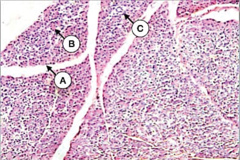
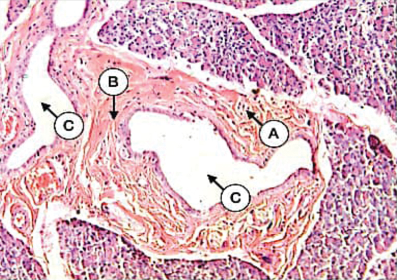
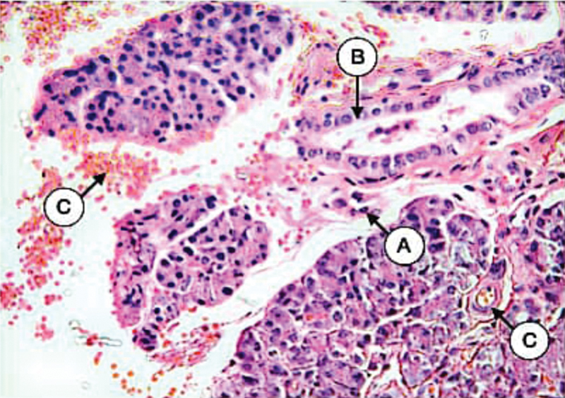
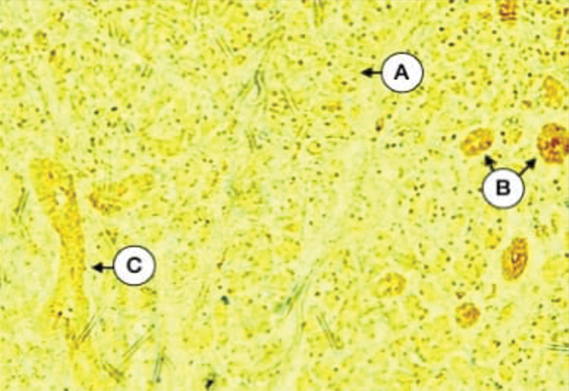
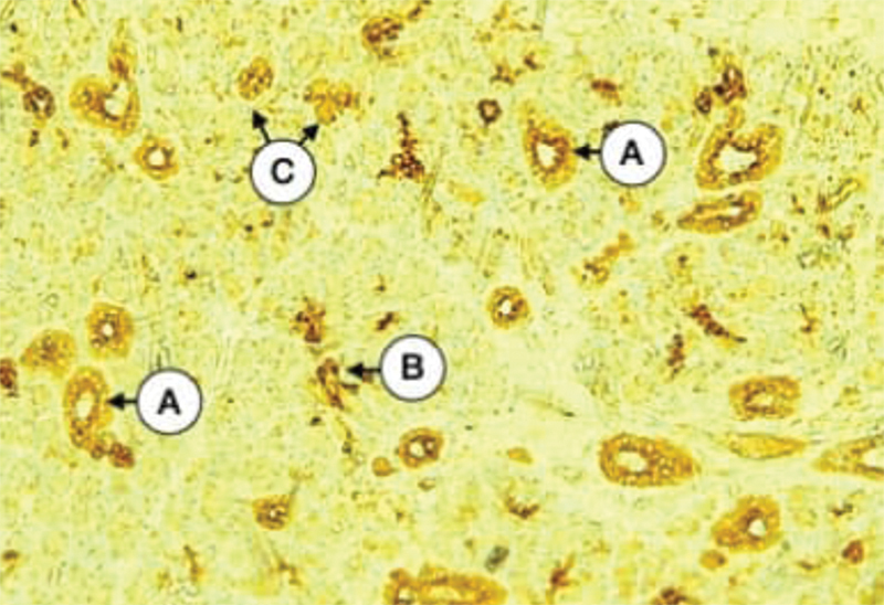
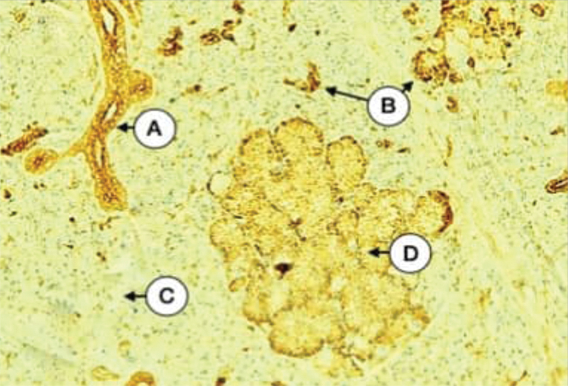
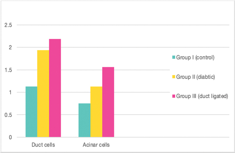
Results: Histological findings confirmed that the parotid gland parenchyma of the diabetic group had glandular atrophy characterized by the loss of gland structure, degenerated acini, and dilatation of the duct system. Moreover, there is a predominance of the fibrous component with the presence of fat cells within the gland compartments. On the contrary, the excretory duct-ligated group undergoes severe glandular atrophy of the previous character with the presence of duct-like structure as well as extravasation and vasodilatation. Immunohistochemical expression of cytokeratin 17 in control parotid using an immunoperoxidase technique showed that cytokeratin expression varies from negative to mild in all ducts and some serous acinar cells. The gland parenchyma of the diabetic group showed mild to strong cytokeratin expression of duct cells more concentrated in the apical part with moderate to strong expression of diffuse type in some serous acini. The intensity of cytokeratin 17 in gland compartments of the excretory duct-ligated group revealed a variation in expression that ranged from negative to strong diffuse staining throughout the gland.
Conclusion: The severity and prevalence of cytokeratin 17 in our results are predictive of the pathological influence of both diabetes mellitus and duct ligation on the cytokeratin intracellular filaments of the salivary gland parenchyma in a different way that interferes with saliva production and/or secretion leading to xerostomia.
The Author(s). This is an open access article published by Thieme under the terms of the Creative Commons Attribution License, permitting unrestricted use, distribution, and reproduction so long as the original work is properly cited. (https://creativecommons.org/licenses/by/4.0/).
Conflict of interest statement
None declared.
Figures
Similar articles
-
Immunohistochemical Evaluation of the Pathological Effects of Diabetes Mellitus on the Major Salivary Glands of Albino Rats.Eur J Dent. 2023 May;17(2):485-491. doi: 10.1055/s-0042-1749159. Epub 2022 Jul 4. Eur J Dent. 2023. PMID: 35785821 Free PMC article.
-
Early Alterations of Cytoskeletal Proteins Induced by Radiation Therapy in the Parenchymal Cells of Rat Major Salivary Glands: A Comparative Immunohistochemical Analysis.Cureus. 2024 Dec 13;16(12):e75634. doi: 10.7759/cureus.75634. eCollection 2024 Dec. Cureus. 2024. PMID: 39803043 Free PMC article.
-
[Changes of myoepithelial cells during regeneration of parotid gland].Hua Xi Kou Qiang Yi Xue Za Zhi. 2014 Oct;32(5):446-9. doi: 10.7518/hxkq.2014.05.005. Hua Xi Kou Qiang Yi Xue Za Zhi. 2014. PMID: 25490819 Free PMC article. Chinese.
-
Immunocytochemistry of myoepithelial cells in the salivary glands.Prog Histochem Cytochem. 2003;38(4):343-426. doi: 10.1016/s0079-6336(03)80001-3. Prog Histochem Cytochem. 2003. PMID: 14509196 Review.
-
Histology of the major salivary glands.Am J Surg Pathol. 1989 Oct;13(10):879-99. doi: 10.1097/00000478-198910000-00008. Am J Surg Pathol. 1989. PMID: 2675654 Review.
Cited by
-
Immunohistochemical Evaluation of the Pathological Effects of Diabetes Mellitus on the Major Salivary Glands of Albino Rats.Eur J Dent. 2023 May;17(2):485-491. doi: 10.1055/s-0042-1749159. Epub 2022 Jul 4. Eur J Dent. 2023. PMID: 35785821 Free PMC article.
-
Long-Term Regenerative Potential of Submandibular Glands in Albino Rats Following Radiotherapy: Role of Cytokeratin 17 Redistribution.Cureus. 2025 Mar 30;17(3):e81465. doi: 10.7759/cureus.81465. eCollection 2025 Mar. Cureus. 2025. PMID: 40303535 Free PMC article.
-
Early Alterations of Cytoskeletal Proteins Induced by Radiation Therapy in the Parenchymal Cells of Rat Major Salivary Glands: A Comparative Immunohistochemical Analysis.Cureus. 2024 Dec 13;16(12):e75634. doi: 10.7759/cureus.75634. eCollection 2024 Dec. Cureus. 2024. PMID: 39803043 Free PMC article.
-
The efficacy of using metformin and/or quercetin for amelioration of gamma-irradiation induced tongue toxicity in diabetic rats.BMC Oral Health. 2024 Jan 18;24(1):110. doi: 10.1186/s12903-024-03871-0. BMC Oral Health. 2024. PMID: 38238729 Free PMC article.
-
BuZhong YiQi Formula Alleviates Diabetes-Caused Hyposalivation by Activating Salivary Secretion Pathway in the Parotid and Submandibular Glands of Rats.Pharmaceuticals (Basel). 2025 Mar 6;18(3):377. doi: 10.3390/ph18030377. Pharmaceuticals (Basel). 2025. PMID: 40143153 Free PMC article.
References
-
- Mahmoud A, Sherif S H. Expression of cytokeratin, actin and proliferating cell nuclear antigen (PCNA) in parotid gland of normal and alloxan-induced diabetic dogs. (Histological and immunohisto-chemical study) Cairo Dent J. 2015;31(02):2661–2674.
-
- Dodds M W, Johnson D A, Yeh C K. Health benefits of saliva: a review. J Dent. 2005;33(03):223–233. - PubMed
-
- Schipper R G, Silletti E, Vingerhoeds M H. Saliva as research material: biochemical, physicochemical and practical aspects. Arch Oral Biol. 2007;52(12):1114–1135. - PubMed
-
- Maria O M, Maria S M, Redman R S et al.Effects of double ligation of Stensen's duct on the rabbit parotid gland. Biotech Histochem. 2014;89(03):181–198. - PubMed
-
- Gaber W, Shalaan S, Misk N, Ibrahim A. Surgical anatomy, morphometry, and histochemistry of major salivary glands in dogs: updates and recommendations. Int J Vet Health Sci Res. 2020;8(02):252–259.
LinkOut - more resources
Full Text Sources