. 2022 Jul;33(7):1402-1410.
doi: 10.1681/ASN.2022010060. Epub 2022 Jun 21.

Hypomagnesemia, Hypocalcemia, and Tubulointerstitial Nephropathy Caused by Claudin-16 Autoantibodies

Affiliations

Hypomagnesemia, Hypocalcemia, and Tubulointerstitial Nephropathy Caused by Claudin-16 Autoantibodies

Lucile Figueres et al. J Am Soc Nephrol. 2022 Jul.

Abstract

Background: Chronic hypomagnesemia is commonly due to diarrhea, alcoholism, and drugs. More rarely, it is caused by genetic defects in the effectors of renal magnesium reabsorption.

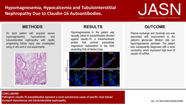
Methods: In an adult patient with acquired severe hypomagnesemia, hypocalcemia, tubulointerstitial nephropathy, and rapidly progressing kidney injury, similarities between the patient's presentation and features of genetic disorders of renal magnesium transport prompted us to investigate whether the patient had an acquired autoimmune cause of renal magnesium wasting. To determine if the patient's condition might be explained by autoantibodies directed against claudin-16 or claudin-19, transmembrane paracellular proteins involved in renal magnesium absorption, we conducted experiments with claudin knockout mice and transfected mouse kidney cells expressing human claudin-16 or claudin-19. We also examined effects on renal magnesium handling in rats given intravenous injections of IgG purified from sera from the patient or controls.

Results: Experiments with the knockout mice and in vitro transfected cells demonstrated that hypomagnesemia in the patient was causally linked to autoantibodies directed against claudin-16, which controls paracellular magnesium reabsorption in the thick ascending limb of Henle's loop. Intravenous injection of IgG purified from the patient's serum induced a marked urinary waste of magnesium in rats. Immunosuppressive treatment combining plasma exchange and rituximab was associated with improvement in the patient's GFR, but hypomagnesemia persisted. The patient was subsequently diagnosed with a renal carcinoma that expressed a high level of claudin-16 mRNA.

Conclusions: Pathogenic claudin-16 autoantibodies represent a novel autoimmune cause of specific renal tubular transport disturbances and tubulointerstitial nephropathy. Screening for autoantibodies targeting claudin-16, and potentially other magnesium transporters or channels in the kidney, may be warranted in patients with acquired unexplained hypomagnesemia.

Keywords: autoantibodies; claudin-16; hypocalcemia; hypomagnesemia.


Figures

None
Graphical abstract
Figure 1.
Figure 1.
The patient’s clinical history. (A) A 71-year-old male patient presented with acquired severe hypomagnesemia, hypocalcemia, and rapidly progressing kidney injury. His SCr, SCa, and SMg levels were normal 18 months earlier. Administration of intravenous Mg chloride induced partial and transient correction of hypomagnesemia, but oral Mg supplementation had no effect on SMg level. SCa level increased to near normal values with oral Ca supplementation. The patient developed rapidly progressing kidney injury. Kidney biopsy disclosed features of subacute or chronic tubular injury (A, light microscopy study, periodic acid–Schiff stain. Original magnification: ×100, bar = 100 µm and Supplemental Figure 1A). After claudin-16 antibodies were detected (see Figures 2 and 3), the patient underwent five sessions of plasma exchange followed by two weekly infusions of rituximab. Renal function gradually improved but hypomagnesemia persisted. Then 3 years after initial presentation, right renal tumor extending to the renal vein was diagnosed (B, asterisk [*] shows enhanced computed tomography scan). Right nephrectomy was performed. Pathologic examination disclosed an undifferentiated grade 4 RCC (Supplemental Figure 1B). After renal tumor removal, the SMg level increased but remained below normal values. Then 9 months after surgery, RCC relapsed (retroperitoneal lymphoadenopathy) (C, asterisk [*] shows enhanced computed tomography scan) and the SMg level decreased again. The patient was started on antityrosine kinase therapy, but subsequently died in the setting of metastatic renal carcinoma. IV, intravenous; PE, plasma exchanges; RTX, rituximab.
Figure 2.
Figure 2.
Immunostaining in the cortex of kidney tissue from mice. (A) Sections of fixed fresh frozen kidney from wild-type mice were incubated with the patient’s plasma (1:500) and rabbit anti-NKCC2 antibody (1:7500) and subsequently with Alexa 555–conjugated goat antihuman IgG (1:600) and Alexa 488–conjugated goat antirabbit (1:500) antibodies, respectively. The patient’s plasma-stained tubular sections expressing NKCC2 in wild-type mice kidney (arrowheads). This staining was similar to that of paracellular proteins in tight junctions of tubular cells. Bar = 20 µm. (B) Sections of fixed fresh frozen kidney from wild-type mice were incubated with control plasma (1:500) and rabbit anti-NKCC2 antibody (1:7500) and subsequently with Alexa 555–conjugated goat antihuman IgG (1:600) and Alexa 488–conjugated goat antirabbit (1:500) antibodies, respectively. No staining was found at the tight junction in the thick ascending limb of Henle’s loop. Bar = 20 µm. (C) Sections of fixed fresh frozen kidney from Cldn16 KO mice were incubated with the patient’s plasma (1:500), and rabbit anti-NKCC2 antibody (1:7500) and subsequently with Alexa 555–conjugated goat antihuman IgG (1:600) and Alexa 488–conjugated goat antirabbit (1:500) antibodies, respectively. The patient’s plasma did not stain tubular sections in Cldn16 KO mice kidney sections. Bar = 20 µm. (D) Serial sections of fixed fresh frozen kidney from wild-type mice were incubated with either the patient’s plasma (1:500) or a mouse monoclonal anti-CLDN16 antibody (1:25) and subsequently Alexa 555–conjugated goat antihuman IgG (1:600) and Alexa 488–conjugated donkey antimouse (1:500) antibodies, respectively. The patient’s plasma-stained tubular sections with the same subcellular localization as the mouse monoclonal claudin-16 antibody. Serial sections had to be used because distinct antigen retrieval conditions had to be used for immunolabeling with the patient’s plasma and with monoclonal antibody. Bar = 20 µm.
Figure 3.
Figure 3.
Staining of transfected immortalized kidney tubular cells. Transfected immortalized mouse kidney TAL (MKTAL) cells expressing V5-tagged human CLDN16 (stained with anti-V5 Ab) (A, D) or Flag-tagged human CLDN19 (stained with anti-Flag Ab) (G, J) were incubated with either the patient’s plasma (B, H) or control plasma (E, K). Only CLDN16-expressing cells were stained with the patient’s plasma (overlays, C, F, I, L). Bar = 10 µm. See also Supplemental Figure 3 for experiments with CLDN10-expressing cells that were not stained with the patient’s plasma.
Figure 4.
Figure 4.
IgG purified from the patient’s serum and from the serum of four patients with chronic tubulointerstitial nephropathy of undetermined cause were injected intravenously in male Sprague-Dawley rats at day 0 and day 2 (n=4, in each group). The injection of the patient’s IgG, but not of control IgG, was associated to a significant increase in renal fractional Mg (FEMg) excretion on day 3 (P=0.03 for patient’s IgG versus control IgG by two-way ANOVA test; P=0.04 for the patient’s IgG day 0 versus day 3 by one-way ANOVA test). Renal calcium and sodium excretion, and serum Mg, calcium and creatinine levels did not vary in both groups of rats (see Supplemental Figure 4).

Comment in

  • Autoimmune Renal Calcium and Magnesium Wasting.
    Schlingmann KP, Konrad M. Schlingmann KP, et al. J Am Soc Nephrol. 2022 Jul;33(7):1231-1233. doi: 10.1681/ASN.2022050545. Epub 2022 Jun 21. J Am Soc Nephrol. 2022. PMID: 35728888 Free PMC article. No abstract available.

References

    1. de Baaij JH, Hoenderop JG, Bindels RJ: Magnesium in man: Implications for health and disease. Physiol Rev 95: 1–46, 2015 - PubMed
    1. Houillier P: Mechanisms and regulation of renal magnesium transport. Annu Rev Physiol 76: 411–430, 2014 - PubMed
    1. Olinger E, Houillier P, Devuyst O: Claudins: A tale of interactions in the thick ascending limb. Kidney Int 93: 535–537, 2018 - PubMed
    1. Agus ZS: Mechanisms and causes of hypomagnesemia. Curr Opin Nephrol Hypertens 25: 301–307, 2016 - PubMed
    1. Alexander RT, Hoenderop JG, Bindels RJ: Molecular determinants of magnesium homeostasis: Insights from human disease. J Am Soc Nephrol 19: 1451–1458, 2008 - PMC - PubMed

Publication types

LinkOut - more resources