. 2022 Dec 1;60(6):2102725.
doi: 10.1183/13993003.02725-2021. Print 2022 Dec.

Human lungs show limited permissiveness for SARS-CoV-2 due to scarce ACE2 levels but virus-induced expansion of inflammatory macrophages

Affiliations

Human lungs show limited permissiveness for SARS-CoV-2 due to scarce ACE2 levels but virus-induced expansion of inflammatory macrophages

Katja Hönzke et al. Eur Respir J. .

Erratum in

Abstract

Background: Severe acute respiratory syndrome coronavirus 2 (SARS-CoV-2) utilises the angiotensin-converting enzyme 2 (ACE2) transmembrane peptidase as cellular entry receptor. However, whether SARS-CoV-2 in the alveolar compartment is strictly ACE2-dependent and to what extent virus-induced tissue damage and/or direct immune activation determines early pathogenesis is still elusive.

Methods: Spectral microscopy, single-cell/-nucleus RNA sequencing or ACE2 "gain-of-function" experiments were applied to infected human lung explants and adult stem cell derived human lung organoids to correlate ACE2 and related host factors with SARS-CoV-2 tropism, propagation, virulence and immune activation compared to SARS-CoV, influenza and Middle East respiratory syndrome coronavirus (MERS-CoV). Coronavirus disease 2019 (COVID-19) autopsy material was used to validate ex vivo results.

Results: We provide evidence that alveolar ACE2 expression must be considered scarce, thereby limiting SARS-CoV-2 propagation and virus-induced tissue damage in the human alveolus. Instead, ex vivo infected human lungs and COVID-19 autopsy samples showed that alveolar macrophages were frequently positive for SARS-CoV-2. Single-cell/-nucleus transcriptomics further revealed nonproductive virus uptake and a related inflammatory and anti-viral activation, especially in "inflammatory alveolar macrophages", comparable to those induced by SARS-CoV and MERS-CoV, but different from NL63 or influenza virus infection.

Conclusions: Collectively, our findings indicate that severe lung injury in COVID-19 probably results from a macrophage-triggered immune activation rather than direct viral damage of the alveolar compartment.


Conflict of interest statement

Conflict of interest: J-C. Rückert and H. Radbruch report support from DFG RA 2491/1-1, BMBF (Defeat Pandemics). A.E. Hauser reports support from Charité – Universitätsmedizin Berlin and Deutsches Rheuma-Forschungszentrum Berlin, and grants from Deutsche Forschungsgemeinschaft (HA5354/10-1, TRR130,P17 and C01, HA5354/8-1). T. Wolff reports support from Federal Ministry of Education and Research (BMBF) grant 01K12006F. M. Kessler reports grants from BMBF Organo-Strat, Einstein 3R. M. Dohmen reports contracts with Max-Delbrück Center, Berlin; grants from Gender Equality Fund, Berlin Institute of Health. F. Klauschen reports consulting fees, lecture honoraria, travel support and participation on advisory boards with BMS, Novartis, Roche and Lilly, and is a co-founder of AI-BIH/Charité-Spinoff Aignostics GmbH. F. Heppner reports consulting fees, lecture honoraria, payment for expert testimony and leadership roles at Novartis, AstraZeneca and ThinkHealth Hygiene Solutions. V.M. Corman reports the following patents: 20210190797 (Methods and reagents for diagnosis of SARS-CoV-2 infection); 9841834 (Human recombinant monoclonal antibody against SARS-CoV-2 spike glycoprotein); 9909654 (A pharmaceutical combination comprising an anti-viral protonophore and a serine protease inhibitor). D. Niemeyer reports that Technische Universität Berlin, Freie Universität Berlin and Charité – Universitätsmedizin have filed a patent application for siRNAs inhibiting SARS-CoV-2 replication with D. Niemeyer as coauthor. M.A. Müller reports the following patents: 20210190797 (Methods and reagents for diagnosis of SARS-CoV-2 infection); 9841834 (Human recombinant monoclonal antibody against SARS-CoV-2 spike glycoprotein); 9909654 (A pharmaceutical combination comprising an anti-viral protonophore and a serine protease inhibitor); and has participated on an advisory board for ECDC/WHO. S. Ludwig reports consulting fees from Atriva Therapeutics GmbH, Biontec SE; and has patent PCT/EP2021/063485 pending. M. Witzenrath reports grants from Deutsche Forschungsgemeinschaft, Bundesministerium für Bildung und Forschung, Deutsche Gesellschaft für Pneumologie, European Respiratory Society, Marie Curie Foundation, Else Kröner Fresenius Stiftung, Capnetz Stiftung, International Max Planck Research School, Quark Pharma, Takeda Pharma, Noxxon, Pantherna, Silence Therapeutics, Vaxxilon, Actelion, Bayer Health Care, Biotest and Boehringer Ingelheim; consulting fees from Noxxon, Pantherna, Silence Therapeutics, Vaxxilon, Aptarion, GlaxoSmithKline, Sinoxa and Biotest; lecture honoraria from AstraZeneca, Berlin Chemie, Chiesi, Novartis, Teva, Actelion, Boehringer Ingelheim, GlaxoSmithKline, Biotest, Bayer Health Care; and has the following patents issued: EPO 12181535.1 (IL-27 for modulation of immune response in acute lung injury), WO/2010/094491 (Means for inhibiting the expression of Ang-2), DE 102020116249.9 (Camostat/Niclosamide cotreatment in SARS-CoV-2 infected human lung cells). All other authors have nothing to disclose.

Figures
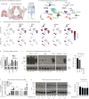
FIGURE 1
FIGURE 1
Angiotensin-converting enzyme 2 (ACE2) expression is scarce in human lungs and cannot be induced by interferon (IFN) at the protein level. a) Schematic illustration of cell types of the investigated alveolar compartment as well as single-cell RNA sequencing (scSeq). b) Annotation of cell clusters from scSeq and single-nucleus RNA sequencing (snSeq) of human lung tissue and autopsy material, respectively. Confirmation of major cell types by related marker gene expression is presented in supplementary figure S1a and b. c) Comparison of scarce ACE2 expression in alveolar epithelial type 2 (AT2) cells with abundant KREMEN1, ASGR1, CD147/BSG, TMPRSS2 and FURIN expression by scSeq and snSeq. d) Analysis of ACE2, CD147/BSG, TMPRSS2 and FURIN expression by quantitative (q)PCR on bulk RNA of normal human lung tissue, human bronchial organoids and Calu-3 cells. Ct values are normalised to glyceraldehyde 3-phosphate dehydrogenase (GAPDH) expression to demonstrate absolute levels of ACE2 compared to other factors. e) Analysis of constitutive ACE2 expression by Western blot using the R&D AF933 antibody. Antibody evaluation is presented in supplementary figure S1c. Shown are protein lysates from human kidneys (three donors), human lungs (five donors), human bronchial organoids (three donors) and Calu-3 cells. Prolonged exposure time (30 s) of membranes was carried out to demonstrate scarce ACE2 expression in lungs and bronchial organoids compared to kidneys and Calu-3. Exposure time of 1 s is shown in supplementary figure S1d. β-Actin served as loading control. Densitometric analysis shows semi-quantitative ACE2 expression normalised to β-actin (right panel). f) Human lung tissue was stimulated for 24 h and 96 h with IFNβ and ACE2, CD147/BSG, TMPRSS2, FURIN and Mx1 expression was analysed by qPCR on bulk RNA. g) Western blot analysis of ACE2 expression in IFNβ-stimulated human lung tissue after 24 h and 96 h of six donors and corresponding densitometric analysis normalised to β-actin. Data are presented as mean±sem. NK: natural killer; UMAP: Uniform Manifold Approximation and Projection. *: p<0.05.
FIGURE 2
FIGURE 2
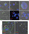
Spectral imaging reveals scarce apical angiotensin-converting enzyme 2 (ACE2) on alveolar epithelial type 2 (AT2) cells, less congruence with ACE2 as well as abundant TMPRSS2 expression in AT1 and AT2. Immunohistochemistry and in situ hybridisation were analysed in human lung tissue and organoids by spectral microscopy and linear unmixing. a) Panel i) shows single-channel detection (to avoid antibody cross-reactivity) of ACE2 immunostaining (green) on the apical surface of an AT2 cell (TII). AT2/ACE2 expression was confirmed by dual-channel detection with AT2 marker HTII-280 (red) in panels iv–vi) (arrows). Panel ii) shows ACE2 mRNA (red) in an AT2 cell. Sequential immunostaining could not reveal ACE2 protein on the apical surface (arrow). Lack of apical ACE2 protein in a fraction of ACE2 mRNA (red) positive cells is demonstrated by lentiviral overexpression in a human bronchial organoid in panel iii). Parts of the cells show apical ACE2 (green) as well. Cell nuclei are visualised by 4′,6-diamidino-2-phenylindole (DAPI) stain (blue). Scale bars: 5 µm (lungs), 10 µm (bronchial organoids). b) Immunostaining for CD68 (alveolar macrophages (AM), orange) and EMP2 (AT1, green) as well as in situ hybridisation for TMPRSS2 (red, panel i)) and HTII-280 (AT2, panel ii), green) in human lung tissue shows TMPRSS2 positive in AT1 (inset) and AT2, but not vessels (V) or AM. Cell nuclei are visualised by DAPI stain (blue). Scale bar: 10 µm. ACE2 expression in Calu-3 cells and kidneys as well as ACE2 antibody tests are presented in supplementary figure S2.
FIGURE 3
FIGURE 3
Severe acute respiratory syndrome coronavirus 2 (SARS-CoV-2) shows low replication on human lung tissue, which can be increased by angiotensin-converting enzyme 2 (ACE2) overexpression. a) Human lung tissue explants (five donors) were infected with human seasonal influenza H3N2 virus A/Panama/2007/1999 (IAV) (orange), Middle East respiratory syndrome coronavirus (MERS-CoV) (blue), SARS-CoV (green) and SARS-CoV-2 (red) with 1×10⁶ plaque-forming units (PFU) and viral replication was assessed. b) Human lung tissue explants (four donors) were infected with IAV H3N2 (orange), MERS-CoV (blue), SARS-CoV (green) with 1×10⁶ PFU and SARS-CoV-2 (red) with 30×106 PFU and viral replication was measured showing validation of figure 3a for all viruses and loss of significance for SARS-CoV-2. External validation experiment of SARS-CoV-2 replication (three donors) is shown in supplementary figure S3a. c) Calu-3 cells were infected with IAV H3N2 (orange), MERS-CoV (blue), SARS-CoV (green) and SARS-CoV-2 (red) with multiplicity of infection (MOI) 0.1 and viral replication was assessed. d) ACE2 expression (Western blot) in wild-type human bronchial organoids (wt), organoids with lentiviral transduction and overexpression of ACE2 (ACE2+) and Calu-3 cells. β-Actin served as loading control. e) ACE2 immunostaining (green) and in situ hybridisation (red) in human ACE2+ bronchial organoids (left panel) showing abundant ACE2 mRNA expression with a sub-fraction of cells positive for apical ACE2 protein expression. The right panel demonstrates a partly apical expression of ACE2 in ACE2+ bronchial organoids as well as a congruency for SARS-CoV-2 S-protein detection in these cells (16 h post-infection, MOI 1). Cell nuclei are visualised by 4′,6-diamidino-2-phenylindole (DAPI) stain (blue). Scale bar=10 µm. f) Wild-type (wt) and ACE2 overexpressing (ACE2+) bronchial organoids (four independent experiments) were infected with SARS-CoV-2 with MOI 1 and viral replication was measured after 0, 16, 24, 48 and 72 h depicting no permissiveness for SARS-CoV-2 when ACE2 is missing and increased permissiveness after ACE2 overexpression. g) Wild-type (wt) alveolar organoids (six independent experiments) were infected with SARS-CoV-2 with MOI 1 and viral replication was measured after 0, 24, 48 and 72 h. h) Uniform Manifold Approximation and Projection embedding of alveolar and bronchial organoid data shows cells positive for SARS-CoV-2 (red). i) ACE2 immunostaining (green, arrows show apical ACE2 expression in SARS-CoV-2 infected cells) and SARS-CoV-2 S-protein detection (red) in human alveolar organoids (24 h post-infection, MOI 1, three independent experiments). Cell nuclei are visualised by DAPI stain (blue). Scale bar: 10 µm. Data are presented as mean±sem. *: p<0.05, **: p<0.01, ***: p<0.001.
FIGURE 4
FIGURE 4
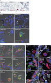
Severe acute respiratory syndrome coronavirus 2 (SARS-CoV-2) has a rare tropism to alveolar epithelial type 2 (AT2), but is frequently phagocytosed by alveolar macrophages (AM) in ex vivo infected human lungs. a) In situ hybridisation for N-gene of SARS-CoV-2 (red) in mock-infected (panel i) and SARS-CoV-2 infected (30×106 plaque-forming units (PFU); panels ii and iii) human lung tissue. Black arrows indicate AT2 cells (TII) and red arrows alveolar macrophages. Cell nuclei are visualised by Hemalaun stain (blue). Scale bars: 50 µm (panels i and ii), 20 µm (panel iii). b) Spectral imaging of immunostained SARS-CoV-2 (N-protein, green) in mock-infected (panel i) and SARS-CoV-2-infected (30×106 PFU; panel ii, iii and iv) human lung tissue confirms rare AT2 tropism, but punctuated viral staining pattern in AM. AT2 cells (TII) and AM are indicated in panels i, ii and iii. Pro-surfactant protein C staining (red) was used in panel iv as AT2 marker. Cell nuclei are visualised by 4′,6-diamidino-2-phenylindole (DAPI) stain (blue). Scale bars: 5 µm (panels i, iii and iv), 10 µm (panel ii). c) Overexpression of angiotensin-converting enzyme 2 (ACE2) (ACE2+, green) in human lung tissue results in broadening of ACE2+ cell types. ACE2-positive AT1 (TI, panel i), endothelial cells (EC, panels i and iv), AT2 (TII, panel ii), bronchial epithelium (BE, panel iii) and AM (panels v and vi) show correlation with SARS-CoV-2 infection (red). Cell nuclei are visualised by DAPI stain (blue). Scale bars: 10 µm. d) Occludin lining and loss (green) indicate virus-induced tissue damage, which is moderate at early and late stages of SARS-CoV-2 infection in human lung tissue (panels i and ii). Overexpression of ACE2 (ACE2+) in human lungs leading to a broadened cellular tropism of SARS-CoV-2 (panel iii) results in an increase of cellular damage at late stages of infection (panel iv), similar to Middle East respiratory syndrome coronavirus (MERS-CoV) (panels v and vi). Cell nuclei are visualised by DAPI stain (blue). Scale bars: 5 µm. MERS-CoV immunostaining and in situ hybridisation in human lung tissue and SARS-CoV-2 immunostaining and in situ hybridisation in Calu-3 cells as well as control staining for ACE2+ and occludin are presented in supplementary figure S4.
FIGURE 5
FIGURE 5
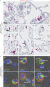
Severe acute respiratory syndrome coronavirus 2 (SARS-CoV-2) detection in alveolar macrophages and fewer alveolar epithelial type 2 (AT2) cells in lungs of an early coronavirus disease 2019 (COVID-19) death. a) In situ hybridisation for N-gene of SARS-CoV-2 (red) in an autopsy lung 5 days after COVID-19 diagnosis. Black arrows indicate AT2 cells and red arrows alveolar macrophages (AM). Cell nuclei are visualised by Hemalaun stain (blue). Scale bars: 50 µm (panel i), 10 µm (panels ii–xi). Additional donors of SARS-CoV-2-positive COVID-19 autopsy lungs and non-COVID-19 autopsy lungs are shown in supplementary figure S5. Infected areas show highly positive cells, but lack of inflammatory cell infiltration indicated by free alveoli and well-shaped alveolar septa. b) Immunolabelling of AT2 cells with HTII-280 (red, TII; panels i, ii and iii), AM with CD68 (red, AM; panels iv, v and vi) and SARS-CoV-2 (green; panels i–vi). Cytosolic staining pattern of AT2 indicate productive infection, whereas the punctuated patterns in AM indicate viral uptake. Note that infected AT2 (TII) detach from the basal membrane (panel ii) and get endocytosed by AM (panels iii and vi). Cell nuclei are visualised by 4′,6-diamidino-2-phenylindole (DAPI) stain (blue). Scale bar: 5 µm. Control staining on autopsy material as well as all other cases are shown in supplementary figure S5.
FIGURE 6
FIGURE 6
Single-cell (scSeq) and single-nucleus (snSeq) RNA sequencing reveals uptake of severe acute respiratory syndrome coronavirus 2 (SARS-CoV-2) by alveolar macrophages and inflammatory response. Human lung tissue explants infected with human seasonal influenza H3N2 virus A/Panama/2007/1999 (IAV) (orange), Middle East respiratory syndrome coronavirus (MERS-CoV) (blue), SARS-CoV (green) at 1×10⁶ plaque-forming units (PFU) and SARS-CoV-2 (red) at 30×106 PFU as outlined in figure 3b was used for scSeq and combined with snSeq data from coronavirus disease 2019 (COVID-19) lung autopsy material. a) scSeq and snSeq of human lung tissue explants and autopsy lungs, respectively; Uniform Manifold Approximation and Projection (UMAP) embedding shows major cell types. b) UMAP embedding shows lung explant cells positive for IAV, MERS-CoV, SARS-CoV or SARS-CoV-2; inset displays SARS-CoV-2-positive alveolar epithelial type 2 (AT2) cells (red) overlaid on angiotensin-converting enzyme 2 (ACE2)-positive cells (blue). c) Quantification of virus-positive cells per cluster (colour key as in a)). Upper panel: comparing results before and after ambient RNA filtering; lower panel: comparing lung explants to lung autopsy material. Error bars represent standard deviation; Pearson correlation values are indicated. d) Quantification of the host factors ACE2, CD147/BSG, NRP1, TMPRSS2, FURIN and DPP4. Expression is averaged for all cells within one cluster of one sample, and then averaged across samples. Error bars represent standard deviation. e) Induction of antiviral and inflammatory pathways: gene expression scores for relevant pathways are averaged across all cells per cluster and condition; z-scores are computed separately for explant and autopsy data. Further analysis is presented in supplementary figure S6.
FIGURE 7
FIGURE 7
Single-cell RNA sequencing reveals inflammatory response of alveolar macrophages (AM) and nonproductive uptake in the absence of angiotensin-converting enzyme 2 (ACE2). Subclustering of macrophages from figure 6, and analysis of isolated lung macrophages infected ex vivo with severe acute respiratory syndrome coronavirus 2 (SARS-CoV-2) (30×106 plaque-forming units (PFU)) and NL63-CoV (1×10⁶ PFU). a) Subclustering of macrophages reveals four subpopulations. Dashed lines serve as a guide to the eye. b) Expression of the top five marker genes for each subcluster in a). c) Gene expression scores for marker-associated pathways from b). Scores are averaged across all cells per subcluster and z-scores are computed separately for explant and autopsy data. d) SARS-CoV-2-positive cells in macrophages from lung tissue explants. e) Compositional changes of macrophage subclusters. p-values from a mixed-effects binomial model. *: p<0.05, **: p<0.01, ***: p<0.001. f) Ex vivo infected lung macrophages. Left panel: percentage of ACE2- or virus-positive cells infected with NL63-CoV or SARS-CoV-2 under mock treatment or ACE2 overexpression, respectively. Right panel: viral reads (as percentage of total) and subgenomic reads (as percentage of viral) for NL63-CoV and SARS-CoV-2, respectively. p-values from binomial model. ***: p<0.001. g) Distribution of reads mapping to the SARS-CoV-2 genome for ex vivo infected AM under mock treatment or ACE2 overexpression. Top panel: coverage per million mapped reads smoothed with 10 nt moving average. Shaded area indicates mean±sem across replicates. Bottom panel: log2 ratio between ACE2 and mock conditions. h) Differential gene expression and pathway analysis for mock-treated ex vivo infected lung macrophages. Left panel: log2 fold change for NL63-CoV versus control plotted against log2 fold change for SARS-CoV-2 versus control. Interferon-stimulated genes (adjusted p<0.01) are highlighted. Right panel: pathway enrichment analysis for the log2 fold changes values with tmod [48]. Further analysis is presented in supplementary figure S7. UMAP: Uniform Manifold Approximation and Projection.

Comment in

References

    1. Bellani G, Laffey JG, Pham T, et al. Epidemiology, patterns of care, and mortality for patients with acute respiratory distress syndrome in intensive care units in 50 countries. JAMA 2016; 315: 788–800. doi: 10.1001/jama.2016.0291 - DOI - PubMed
    1. Cummings MJ, Baldwin MR, Abrams D, et al. Epidemiology, clinical course, and outcomes of critically ill adults with COVID-19 in New York City: a prospective cohort study. Lancet 2020; 395: 1763–1770. doi: 10.1016/S0140-6736(20)31189-2 - DOI - PMC - PubMed
    1. Wu C, Chen X, Cai Y, et al. Risk factors associated with acute respiratory distress syndrome and death in patients with coronavirus disease 2019 pneumonia in Wuhan, China. JAMA Intern Med 2020; 180: 934–943. doi: 10.1001/jamainternmed.2020.0994 - DOI - PMC - PubMed
    1. Ulloa AC, Buchan SA, Daneman N, et al. Estimates of SARS-CoV-2 omicron variant severity in Ontario, Canada. JAMA 2022; 327: 1286–1288. doi: 10.1001/jama.2022.2274 - DOI - PMC - PubMed
    1. Wolter N, Jassat W, Walaza S, et al. Early assessment of the clinical severity of the SARS-CoV-2 omicron variant in South Africa: a data linkage study. Lancet 2022; 399: 437–446. doi: 10.1016/S0140-6736(22)00017-4 - DOI - PMC - PubMed

Publication types

Substances