Review
. 2022 Nov:91:101094.
doi: 10.1016/j.preteyeres.2022.101094. Epub 2022 Jun 18.

Caveolar and non-Caveolar Caveolin-1 in ocular homeostasis and disease

Affiliations
Review

Caveolar and non-Caveolar Caveolin-1 in ocular homeostasis and disease

Eric N Enyong et al. Prog Retin Eye Res. 2022 Nov.

Abstract

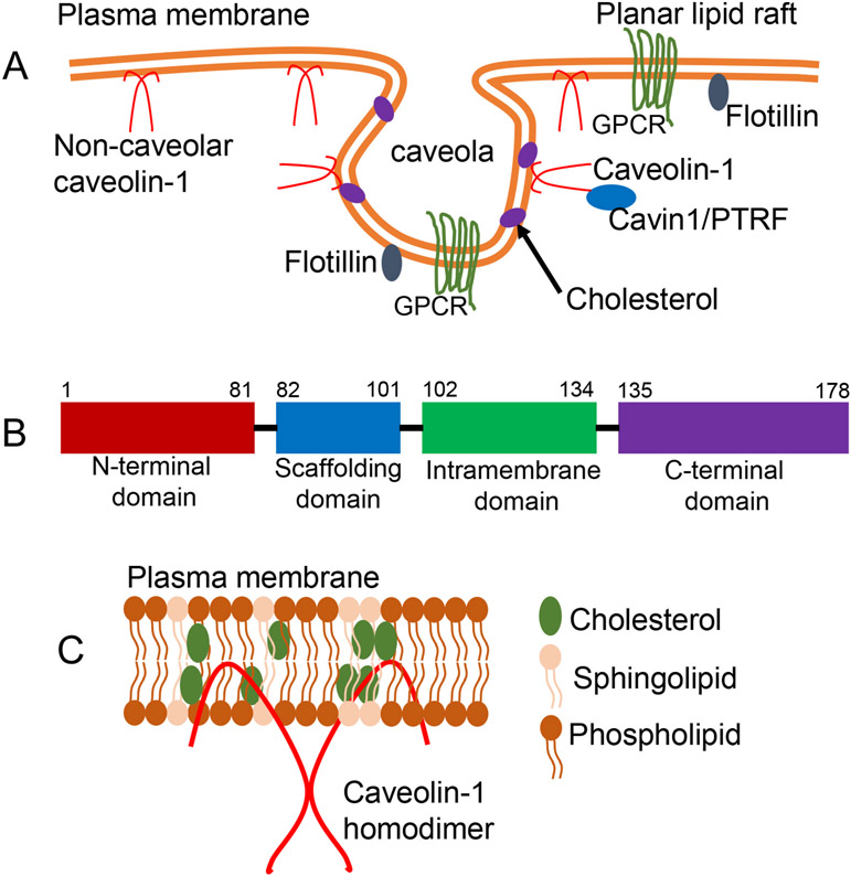
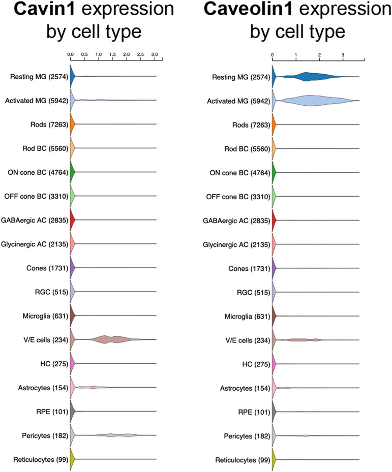
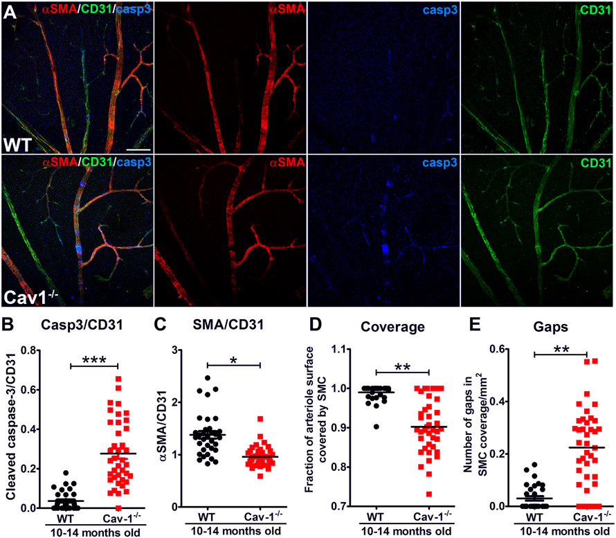
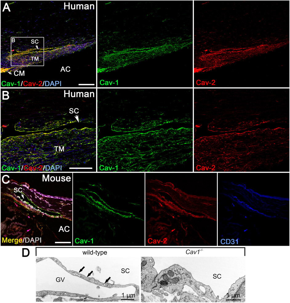
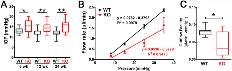
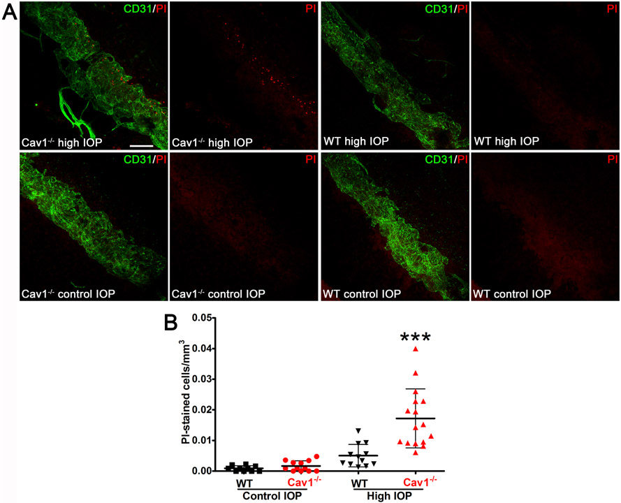
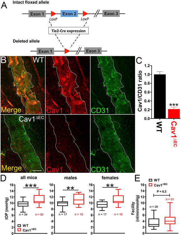
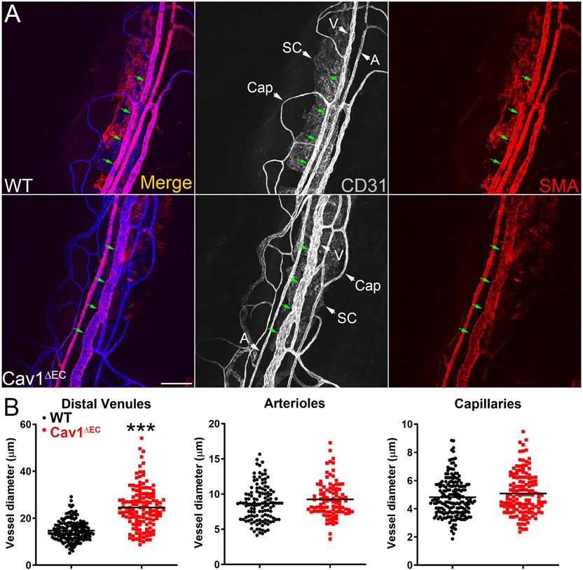
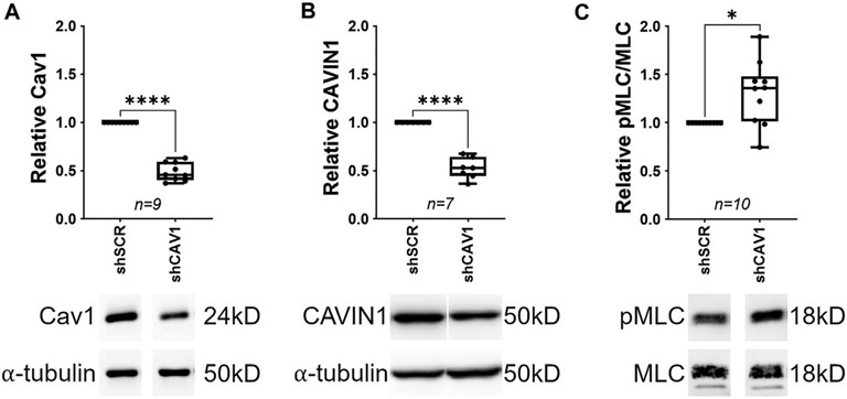
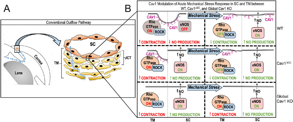
Caveolae, specialized plasma membrane invaginations present in most cell types, play important roles in multiple cellular processes including cell signaling, lipid uptake and metabolism, endocytosis and mechanotransduction. They are found in almost all cell types but most abundant in endothelial cells, adipocytes and fibroblasts. Caveolin-1 (Cav1), the signature structural protein of caveolae was the first protein associated with caveolae, and in association with Cavin1/PTRF is required for caveolae formation. Genetic ablation of either Cav1 or Cavin1/PTRF downregulates expression of the other resulting in loss of caveolae. Studies using Cav1-deficient mouse models have implicated caveolae with human diseases such as cardiomyopathies, lipodystrophies, diabetes and muscular dystrophies. While caveolins and caveolae are extensively studied in extra-ocular settings, their contributions to ocular function and disease pathogenesis are just beginning to be appreciated. Several putative caveolin/caveolae functions are relevant to the eye and Cav1 is highly expressed in retinal vascular and choroidal endothelium, Müller glia, the retinal pigment epithelium (RPE), and the Schlemm's canal endothelium and trabecular meshwork cells. Variants at the CAV1/2 gene locus are associated with risk of primary open angle glaucoma and the high risk HTRA1 variant for age-related macular degeneration is thought to exert its effect through regulation of Cav1 expression. Caveolins also play important roles in modulating retinal neuroinflammation and blood retinal barrier permeability. In this article, we describe the current state of caveolin/caveolae research in the context of ocular function and pathophysiology. Finally, we discuss new evidence showing that retinal Cav1 exists and functions outside caveolae.

Keywords: Caveolae; Caveolin; Cavin1/PTRF; Glaucoma; Inflammation; Müller glia; Non-caveolar Cav1; Retina; Retinal vasculature.


Figures

Fig. 1.
Fig. 1.
Caveolae are flask-shaped plasma membrane invaginations. A) Schematic representation of planar and caveolar membranes. The plasma membranes of many cells consist of planar (flat) or non-planar (invaginated or flasked-shaped) lipid rafts. In cells expressing both Cav1 and Cavin1/PTRF, Cav1 resides and functions predominantly in caveolae domains. On the other hand, when Cav1 is expressed without Cavin1/PTRF such as in prostate cancer (PC3) cells, Cav1 resides outside caveolae in non-caveolar Cav1 scaffolds. B) Schematic diagram of Cav1 domains. All three caveolins possess an N-terminal domain, a scaffolding domain, an intramembrane domain and a C-terminal domain. C) Diagram showing Cav1 membrane topology and homooligomer of Cav1. Cav1 assumes an unusual membrane topology with both its N- and C- terminals facing the cytoplasm.
Fig. 2.
Fig. 2.
Schematic representation depicting the main features of Cavin family of proteins. All Cavins possess two regions of α-helices called HR1 and HR2, which are rich in basic residues, and linked by disordered acidic sequences called DR1, DR2 and DR3.
Fig. 3.
Fig. 3.
Most Cav1 expression in the retina is from the neuroretina. (A) Schematic representation of neuroretinal Cav1 knockout strategy using Cre-lox recombination technology (Chx10-cre/Cav1-floxed mouse model). (B) Representative western blots of whole cell lysate from different tissues. Neuroretinal deletion of Cav1 produced more than 70% reduction in Cav1 expression in whole cell lysate. (C) Densitometry analysis of western blots showing significant reduction in Cav1 protein expression in retina-specific Cav1 KO whole cell lysate. (D) Immunohistochemistry staining of WT and neuroretina-specific Cav1 KO retina sections, stained with anti-Cav1 antibodies and Müller glia-specific marker, anti-glutamine synthetase (GS) showing high expression of Cav1 in Müller glia, which co-localizes with GS. Neuroretinal deletion of Cav1 significantly downregulates Müller glia Cav1 expression, with no effect on Cav1 expression in choroidal and retinal vasculature. Adapted from (Gurley et al., 2020).
Fig. 4.
Fig. 4.
Transcriptional data replotted from microarray data from individual Müller glia from control and Rhodopsin knockout (Rhod KO) mice originally published by (Roesch et al., 2012). In this model rod death peaks at eight weeks (8 w) of age and cone death at 25 weeks (25 w). Cav1 expression is reduced in Müller glia at the peak of rod degeneration but has recovered by the peak of cone degeneration where as Cavin1/PTRF expression shows an opposing expression pattern. Gfap and Glul are indicators for gliosis and Müller glial differentiation status, respectively.
Fig. 5.
Fig. 5.
Violin plots of cell type-specific expression of Cavin1/PTRF and Cav1 in retinal single cell RNAseq data plotted from Hoang et al. (2020) (Hoang et al., 2020). As shown, Cav1 expression is high in resting and activated (by toxic NMDA insult) Müller glia (MG), vascular endothelium (V/E) and pericytes. Cavin1/PTRF is virtually undetectable in resting MG with a small number of cells increasing in expression upon activation. Cavin1/PTRF expression is high in V/E and pericytes. Available interactively at: https://proteinpaint.stjude.org/F/2019.retina.scRNA.html.
Fig. 6.
Fig. 6.
Neuroretinal Cav1 deletion suppresses endotoxin-induced immune response. (A) Deletion of Cav1 globally suppresses inflammatory cytokine secretion into the retina in response to TLR4 activation. (B) Neuroretinal deletion of Cav1 suppresses proinflammatory cytokine secretion into the retina in response to TLR4 activation. In both global and neuroretinal Cav1 KO models, there is a suppression of proinflammatory cytokine secretion into the retina. Proinflammatory cytokines were measured using a multiplex cytokine panel after intraocular injection of LPS. (C) Global Cav1 deletion enhances immune cell influx into the retina. (D) Neuroretina deletion of Cav1 suppresses immune cell influx into the retina. Leukocyte infiltration into the retina was measured by flow cytometry. A and C adapted from (Li et al., 2014); B and D adapted from (Gurley et al., 2020).
Fig. 7.
Fig. 7.
Schematic diagram representing choroidal and intraretinal vascular beds. Diagram illustrates the composition of intraretinal vasculature that supports the inner neural retina (top) and choroidal vasculature that supports the outer neural retina (bottom). The intraretinal vessels form three distinct interconnected (superficial, intermediate, and deep) layers, which are associated with mural SMCs and or PCs. The choroidal vessels and supporting mural SMCs and PCs are located posterior to the RPE and Bruch’s membrane and are composed of the choriocapillaris (CC), Sattler’s layer, and Haller’s layer. Black arrows indicated SMCs on retinal arterioles.
Fig. 8.
Fig. 8.
Endogenous albumin detection in Global-, NR-, and Endo-Cav1 KO retinal tissue. Immunohistochemical staining of mouse retinal sections from global-Cav1 KO (top; adapted from (Gu et al., 2014a)) compared to NR- and Endo-Cav1 KO animals (bottom; (Gurley et al., 2020). Enhanced endogenous albumin detection was observed in global--Cav1 KO retinas and Endo(Tie2)-Cav1 KO animals compared to WT and NR (Chx10)-Cav1 KO retinas. White arrows in the top panel from the Gu et al., 2014a study highlight areas of albumin leakage in global-Cav1 KO superficial vessels. Black arrow in the bottom panel from the Gurley et al. (2020) study indicates albumin extravasation into the RNFL in Endo-Cav1 KO retinas, whereas albumin is contained within vascular lumens of WT and NR-Cav1 KO retinal vessels (bottom white arrows). WT, wild-type; “Cav1-KO” = global-Cav1 KO; NR, neural retinal; Endo, endothelial; Alb/FITC-Alb, fluorescein isothiocyanate-conjugated albumin; Col IV, collagen IV; CAV1, caveolin-1; DAPI, 4′,6-diamidino-2-pheylindole.
Fig. 9.
Fig. 9.
Cav1 deficiency results in segmental loss of alpha smooth muscle actin (αSMA) in retinal arterioles (A,D,E). The segmental gaps in αSMA staining (red) retain immunoreactivity for the endothelial marker CD31 (green) indicating that the vessel is still intact. Regions deficient in αSMA staining show increased immunoreactivity for cleaved caspase-3 (Casp3) indicating localized cell death at these gaps that are deficient in contractile smooth muscle (A,B,C). Reproduced from (Reagan et al., 2018).
Fig. 10.
Fig. 10.
Localization of caveolins to the human (A,B) and mouse (C) conventional outflow tract. Caveolae are abundant features of the Schlemm’s canal (SC) endothelium (D) and trabecular meshwork and are absent from global Cav1−/− mice. Adapted from (Elliott et al., 2016).
Fig. 11.
Fig. 11.
Global Cav1 deficiency results in increased IOP as measured by rebound tonometry (A) and reduced outflow facility (B,C) as measured by ex vivo perfusion. Adapted from (Elliott et al., 2016).
Fig. 12.
Fig. 12.
Caveolae protect conventional outflow pathway tissue against rupture from experimentally-induced IOP elevations. Eyes were pressurized to 50 mmHg for 30 min and tissue was stained with propidium iodide (PI). Cell rupture allows PI to enter cells and label nuclei. As shown in representative panels (A) and in the quantitative analysis (B), caveolae ablation results in enhanced IOP-induced cell rupture. Reproduced from (Elliott et al., 2016).
Fig. 13.
Fig. 13.
Impact of endothelial Cav1 on outflow pathway function. Endothelium-specific Cav1 null mice (Cav1ΔEC) were generated by Cre/lox technology using Cav1 floxed and Tie2-Cre mice (A) resulting in efficient deletion of Cav1 from the SC endothelium (B,C). Cav1ΔEC mice display modest but significant IOP elevation (D) without a concomitant reduction in outflow facility (E). Adapted from (De Ieso et al., 2020).
Fig. 14.
Fig. 14.
Limbal venules (V) downstream of the SC (the distal outflow vessels) are specifically enlarged in Cav1ΔEC mice. Representative confocal images (A) of the limbal region of mouse anterior segment wholemounts stained with Pecam1 (CD31) and alpha smooth muscle actin (SMA). Distal limbal venules (V) but not limbal arterioles (A) or capillaries (Cap) are significantly enlarged in Cav1ΔEC eyes (B). Reproduced from (De Ieso et al., 2020).
Fig. 15.
Fig. 15.
Silencing Cav1 (A) in human trabecular meshwork (TM) cells results in downregulation of Cavin1 (B) and increased phosphorylation of myosin light chain (C), a surrogate marker for enhanced TM cell contractility. Reproduced from (De Ieso et al., 2022).
Fig. 16.
Fig. 16.
Schematic of how caveolae may modulate the physiologic response to mechanical stress in the SC and TM of the conventional outflow pathway function. Cav1 modulates IOP induced eNOS activation in the SC and, potentially, Rho/ROCK signaling in the TM, independently. (WT, wild type; Cav1ΔEC, endothelium-specific Cav1 KO mouse) Reproduced from (De Ieso et al., 2020).
Fig. 17.
Fig. 17.
Cav1 in MIO-M1 cells exists in denaturation-resistant, high molecular weight complexes. (A) Representative Western blots showing expression of Cav1 and Cavin1 in PC3 prostate cancer cells, MIO-M1 Müller glia and retinal microvascular endothelial cells (RMEC). MIO-M1 Müller glia, like endogenous Müller glia abundantly express Cav1. In these cells and PC3 cells, nearly all of Cav1 is found in high molecular weight complexes that are only dissociated upon heating in reducing SDS-PAGE buffer. On the contrary, Cav1 in RMEC cells is predominantly monomeric both without and with heating in reducing SDS-PAGE sample buffer. (B) Stained gel and representative Western blot showing that the high molecular Cav1 complexes are recoverable after immunoprecipitation. High molecular weight complex bands from the stained gel (B1, B2 and B3 indicated by arrows) were excised and analyzed by mass spectrometry. Adapted from Enyong et al., 2022) (Enyong et al., 2022).
Fig. 18.
Fig. 18.
Proteins that interact with high molecular weight Cav1 complexes are associated with the cell cytoskeleton. Cav1 protein-protein interaction by STRING analysis. Cav1 complexes were analyzed by mass spectrometry and STRING open-source database was used to identify protein-protein interactions. A total of 33 proteins were found to interact with Cav1 complexes, most of which play a role in the cytoskeletal architecture.

Similar articles

Cited by

References

    1. Abbasi M, Gupta VK, Chitranshi N, Gupta V, Ranjbaran R, Rajput R, Pushpitha K, Kb D, You Y, Salekdeh GH, Parton RG, Mirzaei M, Graham SL, 2021. Inner retinal injury in experimental glaucoma is prevented upon AAV mediated Shp2 silencing in a caveolin dependent manner. Theranostics 11, 6154–6172. - PMC - PubMed
    1. Abbasi M, Gupta VK, Chitranshi N, Gupta VB, Mirzaei M, Dheer Y, Garthwaite L, Zaw T, Parton RG, You Y, Graham SL, 2020. Caveolin-1 ablation imparts partial protection against inner retinal injury in experimental glaucoma and reduces apoptotic activation. Mol. Neurobiol 57, 3759–3784. - PubMed
    1. Aboulaich N, Vainonen JP, Stralfors P, Vener AV, 2004. Vectorial proteomics reveal targeting, phosphorylation and specific fragmentation of polymerase I and transcript release factor (PTRF) at the surface of caveolae in human adipocytes. Biochem. J 383, 237–248. - PMC - PubMed
    1. Aga M, Bradley JM, Wanchu R, Yang YF, Acott TS, Keller KE, 2014. Differential effects of caveolin-1 and -2 knockdown on aqueous outflow and altered extracellular matrix turnover in caveolin-silenced trabecular meshwork cells. Invest. Ophthalmol. Vis. Sci 55, 5497–5509. - PMC - PubMed
    1. Aghsaei Fard M, Ritch R, 2020. Optical coherence tomography angiography in glaucoma. Ann. Transl. Med 8, 1204. - PMC - PubMed

Publication types

Substances