Review
. 2023 Jan;33(1):30-47.
doi: 10.1016/j.tcb.2022.05.004. Epub 2022 Jun 18.

J-domain protein chaperone circuits in proteostasis and disease

Affiliations
Review

J-domain protein chaperone circuits in proteostasis and disease

Ruobing Zhang et al. Trends Cell Biol. 2023 Jan.

Abstract

The J-domain proteins (JDP) form the largest protein family among cellular chaperones. In cooperation with the Hsp70 chaperone system, these co-chaperones orchestrate a plethora of distinct functions, including those that help maintain cellular proteostasis and development. JDPs evolved largely through the fusion of a J-domain with other protein subdomains. The highly conserved J-domain facilitates the binding and activation of Hsp70s. How JDPs (re)wire Hsp70 chaperone circuits and promote functional diversity remains insufficiently explained. Here, we discuss recent advances in our understanding of the JDP family with a focus on the regulation built around J-domains to ensure correct pairing and assembly of JDP-Hsp70 machineries that operate on different clientele under various cellular growth conditions.

Keywords: Hsp40; Hsp70; J-domain; J-domain proteins; protein conformational diseases; protein homeostasis.


Conflict of interest statement

Declaration of interests The authors declare that they have no competing interests.

Figures
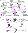
Figure 1.
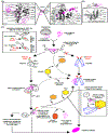
Figure 1.. Selection of client proteins by J-domain proteins for different Hsp70 chaperone functions.
(A) Client selection by JDPs to perform housekeeping and stress-related activities in cells. Shown examples include promiscuous and selective client bindings, as well as JDP mechanisms that do not involve direct client binding. The J-domain is represented as a blue filled circle and dash. The client protein is shown in magenta. (B) Schematic view of the canonical Hsp70 functional cycle. Client proteins are selected and handed over to Hsp70 bound to ATP (nucleotide binding domain (NBD) in dark grey; substrate binding domain (SBD) in light grey). The physical interaction between JDP and Hsp70 is mediated by the J-domain (JD, filled blue circle and dash). JD and client binding synergistically trigger Hsp70’s ATP hydrolysis, resulting in conformational changes in the SBD, which has a high affinity (ADP state) for the client. Subsequently, nucleotide exchange factors (NEF) induce ADP dissociation and re-binding of ATP, converts Hsp70 to low-affinity ATP state. This facilitates the release of the client protein. The released client can fold to its native state or, alternatively, re-enter the Hsp70 cycle.
Figure 2.
Figure 2.. J-domain mediated activation of Hsp70.
(A) Structural view of the J-domain (PDB ID: 1XBL). Helices I-IV are highlighted, as well as the highly conserved His-Pro-Asp (HPD) motif, crucial for biding and stimulating ATPase activity in Hsp70s. (B) Structure of the complex formed between the J-domain (JD, blue) of bacterial JDP (DnaJ) and Hsp70 (DnaK) (PDB ID: 5NRO). Inset panels highlight key residues that form the interaction interface between Hsp70 and the JD (helix II, helix III and HPD loop region). (C) Regulation of Hsp70 ATPase activity by JD and client binding. JD, Hsp70, and client are indicated in blue, grey, and black, respectively. Scenario 1: Binding of ATP induces the two NBD lobes of Hsp70 to rotate into a conformation that keeps the catalytic residues in an ATP hydrolysis-incompetent conformation. This confirmation allows SBDβ to dock onto the lobe-rotated NBD, which is stabilized through the interaction of residues D481 with I168 and K414 with D326. The dissociation of SBDβ from NBD in Hsp70 is infrequent in the absence of JDP and client binding, resulting in a low basal ATP hydrolysis rate. Scenario 2: Client binding can stimulate the dissociation of the SBDβ from the NBD, leading to enhanced ATP hydrolysis in Hsp70, but the linker often slips out of the lower crevice, failing to arrest the lobes in the competent conformation for ATP hydrolysis. This results in a marginal increase in ATPase activity that could prematurely release the client. Scenario 3: The binding of JD prevents the slipping of the linker from the lower crevice and increases the efficiency of the client-stimulated dissociation of SBDβ from NBD. This arrests the back-rotating NBD lobes in the ATP hydrolysis competent conformation, leading to efficient trapping of the client protein. (D) JDP-Hsp70 interaction network. The interaction data are extracted from the BioGrid database, displaying only interactions detected at the physical level. The red connection lines represent interactions that are supported by direct in vitro experimental evidence (“Reconstituted Complex” in BioGrid). ‘Atypical’ denotes putative Hsp70 chaperones with a non-canonical domain architecture (e.g. only the NBD present). The node sizes are scaled by the number of interactions reported for each JDP and Hsp70. Chaperones that are predominantly found in the ER and mitochondria are highlighted by red and green circles, respectively. The rest of the chaperones are located in the cytosol/nucleus.
Figure 3.
Figure 3.. Characteristics of J-domain protein classes.
(A) Classifications of the JDP repertoires by class for H. sapiens, S. cerevisiae, and E. coli. The JDPs found predominantly in the ER and mitochondria are indicated by red and green circles, respectively. The rest of the chaperones are located in the cytosol/nucleus. (*) Proposed reclassification of some class B human JDPs as class C members according to Malinverni et al. [39] (B) Canonical domain architectures and structural view of class A JDPs. (C) Canonical domain architectures and structural view of class B JDPs. (D) Structural views of the nine most abundant JDP architectures in class C. The J-domains (JD) are colored blue. The models shown are full-length predictions obtained by AlphaFold2 as deposited in the EBI database [83]. Below each structure, we show the corresponding simplified domain architecture based on the PFAM domain annotations (excluding the J-domain (PFAM ID: DnaJ) for clarity). Examples of JDPs with the corresponding domain organization in H. sapiens (H.s.), S. cerevisiae (S.c.), and E. coli (E.c.) (when available) are also reported below each example structure. For DUF3444, which is found only in plants, we report an example of a JDP from A. thaliana.
Figure 4.
Figure 4.. Regulation of the J-domain protein-Hsp70 network through the J-domain.
(A) Distribution of exposed electrostatic potentials (iso-surfaces shown at +/− 1kcal/mol, blue: positive potential, red: negative potential) in the J-domains of 50 human JDPs. These electrostatic potential patterns and post-translational modifications could fine-tune the selection of Hsp70. In the absence of structural models, the J-domains were modelled by AlphaFold2. (B) Inhibition of the J-domain of CbpA (blue) by binding of CpbM (orange) (PDB ID 3UCS). The inset displays the critical residues involved in the JD-CbpM interaction interface. (C) Regulation of the J-domain by interactions with the G/F-rich region. Left: Interaction of the yeast Sis1 J-domain with its G/F-rich region through the salt bridge E50-R73 (PDB ID 6D6X). Right: Interaction between helix V and the J-domain of human DNAJB6 (PDB ID 6U3S). (D) Auto-inhibition of human DNAJB8 through the interaction of its J-domain with residues in the C-terminal domain (CTD) (Model provided by L. Joachimiak, Ph.D.).
Box I Figure I.
Box I Figure I.. Client binding and transfer from J-domain proteins to downstream chaperone systems.
(A) Structural view of the interaction between PhoA (unstructured client protein) and CTDs of T. thermophilus DnaJ (PDB ID 6PSI) [14]. The DnaJ residues (grey) interacting with PhoA (pink) are highlighted on the CTDI (left zoom) and CTDII (right zoom). The grey spheres represent C-alpha atoms, and the black spheres represent C-beta atoms. (B) Structural view of the Isu1 binding site at the C-terminus of Jac1 of S. cerevisiae. Zoomed out inset shows residues interacting with Isu1 (PDB ID 3UO2) [114]. Isu1 is a scaffolding protein for the de novo synthesis of iron-sulfur clusters within mitochondria. (C) JDP-mediated client handover pathways to various downstream chaperone systems. The presence of an isolucine vs. a methionine upstream of the EEVD motif allows selective client (magenta) transfer between specific JDPs (e.g. class B) and Hsp70s. This difference can further differentially regulate interactions with Hsp90 (see inset). On the contrary, binding of NudC to class A and B JDPs could dislodge Hsp70 and facilitate the direct transfer of some clients to Hsp90. This bypasses the downstream client transfer between the two chaperone systems via the tetratricopeptide repeat (TPR) domain containing proteins (e.g. HOP and Cdc37). The inset shows how HOP scaffolds Hsp70 and Hsp90 to facilitate client transfer. The interaction of Hsp70 with E3 ubiquitin ligases such as CHIP through the EEVD motif could result in the ubiquitylation and degradation of clients that cannot be efficiently chaperoned.

References

    1. Labbadia J and Morimoto RI (2015) The biology of proteostasis in aging and disease. Annu. Rev. Biochem 84, 435–464 - PMC - PubMed
    1. Hipp MS, et al. (2019) The proteostasis network and its decline in ageing. Nat Rev Mol Cell Biol 20, 421–435 - PubMed
    1. Sui X, et al. (2020) Widespread remodeling of proteome solubility in response to different protein homeostasis stresses. Proc. Natl. Acad. Sci. U. S. A 117, 2422–2431 - PMC - PubMed
    1. Zhao L, et al. (2019) The Hsp70 Chaperone System Stabilizes a Thermo-sensitive Subproteome in E. coli. Cell Rep. 28, 1335–1345.e1336 - PubMed
    1. Ichino L, et al. (2021) MBD5 and MBD6 couple DNA methylation to gene silencing through the J-domain protein SILENZIO. Science 372, 1434–1439 - PMC - PubMed

Publication types

MeSH terms

Substances