. 2022 Jun 22;7(1):192.
doi: 10.1038/s41392-022-01017-8.

Dietary folate drives methionine metabolism to promote cancer development by stabilizing MAT IIA

Affiliations

Dietary folate drives methionine metabolism to promote cancer development by stabilizing MAT IIA

Jin-Tao Li et al. Signal Transduct Target Ther. .

Erratum in

Abstract

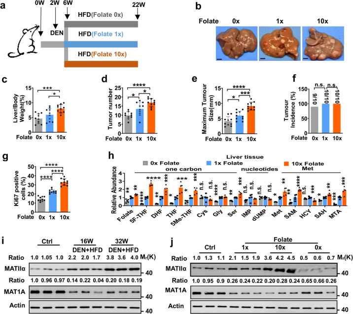
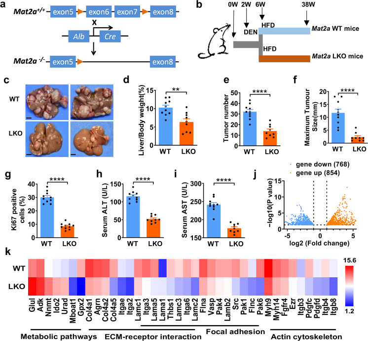
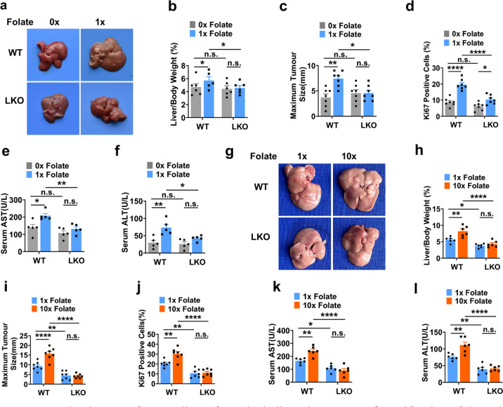
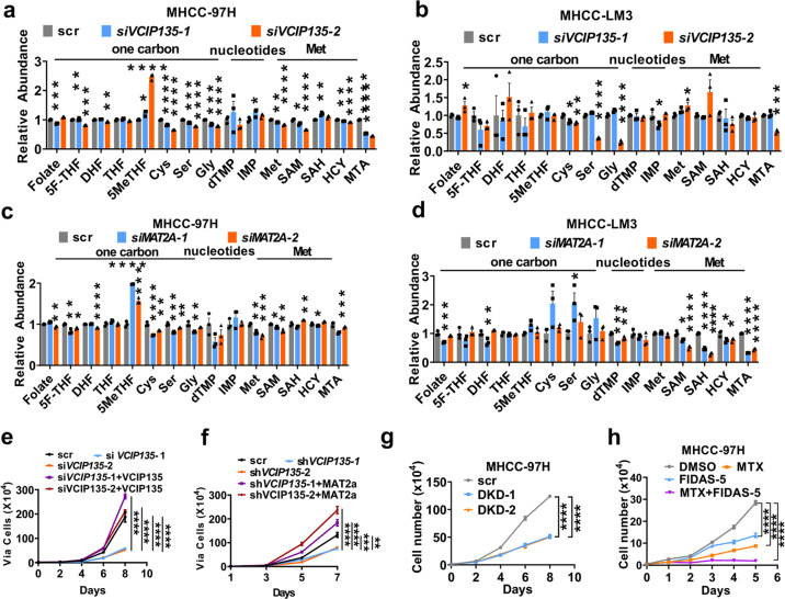
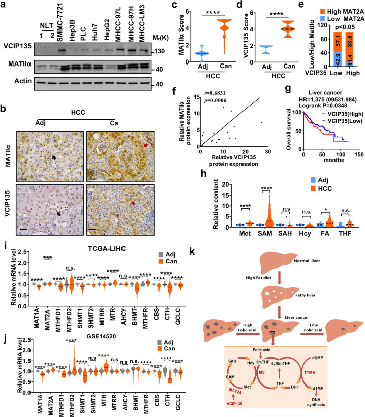
Folic acid, served as dietary supplement, is closely linked to one-carbon metabolism and methionine metabolism. Previous clinical evidence indicated that folic acid supplementation displays dual effect on cancer development, promoting or suppressing tumor formation and progression. However, the underlying mechanism remains to be uncovered. Here, we report that high-folate diet significantly promotes cancer development in mice with hepatocellular carcinoma (HCC) induced by DEN/high-fat diet (HFD), simultaneously with increased expression of methionine adenosyltransferase 2A (gene name, MAT2A; protein name, MATIIα), the key enzyme in methionine metabolism, and acceleration of methionine cycle in cancer tissues. In contrast, folate-free diet reduces MATIIα expression and impedes HFD-induced HCC development. Notably, methionine metabolism is dynamically reprogrammed with valosin-containing protein p97/p47 complex-interacting protein (VCIP135) which functions as a deubiquitylating enzyme to bind and stabilize MATIIα in response to folic acid signal. Consistently, upregulation of MATIIα expression is positively correlated with increased VCIP135 protein level in human HCC tissues compared to adjacent tissues. Furthermore, liver-specific knockout of Mat2a remarkably abolishes the advocating effect of folic acid on HFD-induced HCC, demonstrating that the effect of high or free folate-diet on HFD-induced HCC relies on Mat2a. Moreover, folate and multiple intermediate metabolites in one-carbon metabolism are significantly decreased in vivo and in vitro upon Mat2a deletion. Together, folate promotes the integration of methionine and one-carbon metabolism, contributing to HCC development via hijacking MATIIα metabolic pathway. This study provides insight into folate-promoted cancer development, strongly recommending the tailor-made folate supplement guideline for both sub-healthy populations and patients with cancer expressing high level of MATIIα expression.


Conflict of interest statement

The authors declare no competing interests. Q.-Y.L. is the editorial board member of Signal Transduction and Targeted Therapy, but she has not been involved in the process of the manuscript handling.

Figures

Fig. 1
Fig. 1
MATIIα positively correlates with folic acid-promoted HCC development. a Schematic representation of DEN/HFD-induced HCC model. b Representative images from 22-week-old DEN/HFD-treated mice fed with different concentration folate diet as indicated. ce Quantification of the ratio of liver weight to body weight (c), the number of tumors (d), and the size of the largest tumor per mouse (e). Mean ± s.e.m. of n = 10 biologically independent experiments, one-way ANOVA test. f Quantification of tumor incidence in mice fed with different folate acid diet. Mean ± s.e.m., one-way ANOVA test. g Quantification of Ki67 IHC staining of mice liver tissues. Ten fields each, mean ± s.e.m., one-way ANOVA test. h Quantification of different metabolites of liver tissue in mice fed with different folate diets. Mean ± s.e.m. of n = 5 biologically independent experiments, one-way ANOVA test. i, j Immunoblots of MatIIα and Mat1A in liver tissues from indicated mice. WT mice was employed as Ctrl (control) mice. Data in i and j are representative of three independent experiments. n.s. donates for no significance, *P < 0.05, **P < 0.01, ***P < 0.001 and ****P < 0.0001
Fig. 2
Fig. 2
DUB VCIP135 stabilizes MATIIα to promote cell proliferation by sensesing folic acid. a VCIP135 endogenously interacts with MATIIα. b VCIP135 knockdown increases MATIIα ubiquitylation. HEK293T cells were transfected with the indicated plasmids. c VCIP135 WT but not enzymatic-dead mutant C218S decreases MATIIα ubiquitylation. HEK293T cells were transfected with the indicated plasmids. d, e VCIP135 WT but not enzymatic-dead mutant stabilizes MATIIα upon folate deprivation for 72 h. MHCC-97H cells were cultured under folate-deprived condition, followed by CHX treatment. Mean ± s.d. of n = 3 (d) biologically independent experiments, one-way ANOVA test. f MG132 rescues MTX-reduced MATIIα protein level in multiple HCC cell lines. g Folate rescues MTX-reduced MATIIα protein level in MHCC-97H and Huh7 cells. h Working model. VCIP135 deubiquitylates MATIIα in response to folate and induces MATIIα accumulation in protein level. Data (ac and eg) are representative of three independent experiments. n.s. donates for no significance, ***P < 0.001 and ****P < 0.0001
Fig. 3
Fig. 3
MATIIα is crucial for high fat-induced liver cancer progression. a Generation of liver tissue-specific Mat2a-knockout mice. LoxP was indicated as arrowhead. b Schematic representation of DEN/HFD-induced HCC model. c Representative images of mouse livers from the indicated group. n = 10 (WT), n = 9 (LKO) biologically independent experiments. Scale bar, 5 mm. Quantification of the ratio of liver weight to body weight (d) and the number of tumors (e), quantification of size of the largest tumor per mouse (f). Mean ± s.e.m. of n = 10 (WT), n = 9 (LKO) biologically independent experiments, two-tailed t-test. g Quantification of Ki67 staining of mice liver tissues. Ten fields each, mean ± s.e.m., two-tailed t-test. h, i ALT and AST contents in mice serum. Mean ± s.e.m. of n = 0 (WT), n = 9 (LKO) biologically independent experiments, two-tailed t-test. j Volcano map shows the altered genes and pathways in mice liver via RNA-seq after Mat2a specific knockout in the hepatocytes with 16-week DEN/HFD treatment. k Gene expression levels of the subsets of genes with significant changes in the metabolic pathways, ECM-receptor interaction, Focal adhesion, Actin cytoskeleton in LKO mice at 16 weeks. n = 4 in each group. **P < 0.01, and ****P < 0.0001
Fig. 4
Fig. 4
MATIIα is essential for folic acid-promoted cancer development. a Representative images of mouse livers from the indicated group. b, c Quantification of the ratio of liver weight to body weight (b) and the size of the largest tumor per mouse (c) fed with folate free diet. Mean ± s.e.m. of n = 5–7 biologically independent experiments, one-way ANOVA test. d Quantification of Ki67 staining of mice liver tissues. Seven fields each, mean ± s.e.m., one-way ANOVA test. e, f Serum contents of AST (e) and ALT (f) contents in mice that fed with indicated folate diet. Mean ± s.e.m. of n = 5 biologically independent experiments, one-way ANOVA test. g Representative images of mouse livers from the indicated group. h, i Quantification of the ratio of liver weight to body weight (h) and the size of the largest tumor per mouse (i) fed with folate free diet. Mean ± s.e.m. of n = 5–7 biologically independent experiments, one-way ANOVA test. j Quantification of Ki67 staining of mice liver tissues. Seven fields each, mean ± s.e.m., one-way ANOVA test. k, l Serum contents of AST (k) and ALT (l) contents in mice that fed with indicated folate diet. Mean ± s.e.m. of n = 6 biologically independent experiments, one-way ANOVA test. n.s. donates for no significance, *P < 0.05, **P < 0.01, and ****P < 0.0001
Fig. 5
Fig. 5. MATIIα and VCIP135 are essential for HCC proliferation.
a, b Quantification of different metabolites of VCIP135 knockdown in MHCC-97H cells (a) and MHCC-LM3 cells (b). Mean ± s.e.m. of n = 3 biologically independent experiments, one-way ANOVA test. c, d Quantification of different metabolites of MAT2A knockdown in MHCC-97H cells (c) and MHCC-LM3 cells (d). Mean ± s.e.m. of n = 3 biologically independent experiments, one-way ANOVA test. e VCIP135 putback recovers VCIP135 knockdown-induced reduction of cell proliferation in MHCC-LM3 cells. Mean ± s.e.m. of n = 3 biologically independent experiments, one-way ANOVA test. f MAT2A putback recovers VCIP135 knockdown-induced reduction of cell proliferation in MHCC-LM3 cells in normal medium. Mean ± s.e.m. of n = 3 biologically independent experiments, one-way ANOVA test. g The effects of MAT2A and VCIP135 double knockdown on proliferation in vitro. Mean ± s.e.m. of n = 3 biologically independent experiments, one-way ANOVA test. h MTX and FIDAS-5 synergistically repress cell proliferation. Mean ± s.d. of n = 3 biologically independent experiments, one-way ANOVA test. n.s. donates for no significance, *P < 0.05, **P < 0.01, ***P < 0.001 and ****P < 0.0001
Fig. 6
Fig. 6
MATIIα and VCIP135 are upregulated in HCC and significantly associated with poor prognosis. a VCIP135 and MATIIα protein levels were detected by immunoblotting from HCC cell lines. b Represent images of IHC staining with antibodies against MATIIα or VCIP135 from human HCC patients (n = 58). c, d Quantification of MATIIα (c) and VCIP135 (d) protein levels according to IHC scores in HCC (n = 58). Adj, adjacent; Can, Cancer. Median indicated as dotted lines, two-tailed t-test. e Positive correlation of VCIP135 with MATIIα protein levels determined by IHC staining. The 58 samples were classified into two groups (High VCIP135, 51; Low VCIP135, 7) based on the VCIP135 level. f Positive correlation of VCIP135 with MATIIα protein levels in human hepatocellular cancer tissues samples. VCIP135 and MAT2A protein expression were detected by immunoblotting (n = 21). Spearman correlation, two-tailed. g Kaplan–Meier survival curves for patients with high VCIP135 expression have poorer overall survival compared with patients with low VCIP135 expression in liver cancer. Cutoff value used in analysis is 706. Log-rank (Mantel-Cox) test. FA, folic acid. h Levels of one-carbon metabolites from paired HCCs and non-tumor liver tissues of patients, (n = 44 per group). Median indicated as dotted lines, two-tailed t-test. i, j Different one-carbon metabolism-related genes expression levels in patients (TCGA, i and GSE14520, j). Median indicated as dotted lines, two-tailed t-test. k Working model. Higher folate diet enhances while folate free diet attenuates HCC development. Data in a is representative of three independent experiments. n.s. donates for no significance, *P < 0.05, **P < 0.01, ***P < 0.001 and ****P < 0.0001

References

    1. Sanderson SM, Gao X, Dai Z, Locasale JW. Methionine metabolism in health and cancer: A nexus of diet and precision medicine. Nat. Rev. Cancer. 2019;19:625–637. doi: 10.1038/s41568-019-0187-8. - DOI - PubMed
    1. Mazat JP. One-carbon metabolism in cancer cells: A critical review based on a core model of central metabolism. Biochem. Soc. Trans. 2021;49:1–15. doi: 10.1042/BST20190008. - DOI - PubMed
    1. Sakata SF, Shelly LL, Ruppert S, Schutz G, Chou JY. Cloning and expression of murine S-adenosylmethionine synthetase. J. Biol. Chem. 1993;268:13978–13986. doi: 10.1016/S0021-9258(19)85198-0. - DOI - PubMed
    1. Sakata SF, et al. Effect of fasting on methionine adenosyltransferase expression and the methionine cycle in the mouse liver. J. Nutr. Sci. Vitaminol. 2005;51:118–123. doi: 10.3177/jnsv.51.118. - DOI - PubMed
    1. Lu SC, Mato JM. S-adenosylmethionine in liver health, injury, and cancer. Physiol. Rev. 2012;92:1515–1542. doi: 10.1152/physrev.00047.2011. - DOI - PMC - PubMed

Publication types

MeSH terms