Review
. 2022 Dec;10(2):317-335.
doi: 10.1007/s40487-022-00201-8. Epub 2022 Jun 21.

Basal Cell Carcinoma: A Narrative Review on Contemporary Diagnosis and Management

Affiliations
Review

Basal Cell Carcinoma: A Narrative Review on Contemporary Diagnosis and Management

Piyu Parth Naik et al. Oncol Ther. 2022 Dec.

Abstract

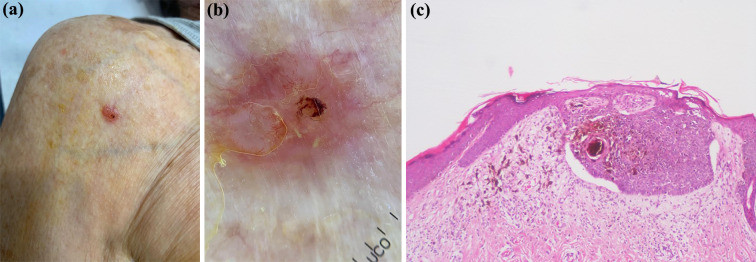
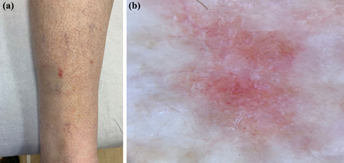
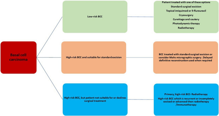
Basal cell carcinoma (BCC) is the most common, accounting for 80-90% of skin cancers. It arises from the basal layer of the epidermis and its appendages. A complex interplay of environmental, phenotypic and genetic variables leads to the development of BCC. Literature has documented several clinical subtypes of BCC, the most common of which are nodular, superficial and morpheaform. Expeditious diagnosis and analysis are essential for improving the outcome of BCC. Preventive measures, particularly when implemented in childhood and adolescence, may play a critical role. Due to its low metastatic potential, treatment for BCC mostly focuses on local management. The standard treatment of basal cell carcinoma involved complete removal of the lesion by excision or Mohs surgery. In special circumstances, basal cell carcinoma can be treated with cryosurgery, electrodesiccation and curettage, topical medications and photodynamic therapy. This review aimed to evaluate the contemporary diagnosis and management of basal cell carcinoma.

Keywords: Basal cell carcinoma; Diagnosis; Management; Surgical excision.


Figures

Fig. 1
Fig. 1
a Clinical photograph of a 42-year-old male patient with a pink-colored, slowly growing nodule on the left forearm for 1 year. b Dermoscopy revealed arborizing telangiectasia. No gray-blue ovoid nests or globules were seen. The provisional clinical diagnosis was basal cell carcinoma. Elliptical excision biopsy with a 3-mm free margin was done under local anesthesia after obtaining written informed consent from the patient. The histopathology report suggested complete excision of the basal cell carcinoma. c Histopathology examination revealed basaloid epithelial cells in the epidermis, typically formed palisades with cleft formation in the basaloid epithelium and crowded nuclei with scattered mitotic figures. The nests remain confined to the papillary dermis, and the lesion seemed to be completely excised from all margins. No lymphatic or perineural invasion was seen. The diagnosis was superficial basal cell carcinoma with free margins, and the lesion was excised completely (H&E staining with  × 10 magnification)
Fig. 2
Fig. 2
a Clinical photograph of a 47-year-old female patient with a pinkish brown, slowly growing erythematous plaque over the right post-auricular upper neck region. The patient reported a gradual change in its size and color over the last 1 year; it had apparently become darker. b Dermoscopy revealed an asymmetrical pattern with irregular margins and arborizing tree-like telangiectasis in the central and peripheral areas. The provisional diagnosis was basal cell carcinoma. Elliptical excisional biopsy under local anesthesia with a 4-mm free margin was done after obtaining all consents. The histopathology report was suggestive of completely excised nodular basal cell carcinoma
Fig. 3
Fig. 3
a Clinical photograph of a 70-year-old female patient with a variable pinkish-colored, slowly growing erythematous plaque over the back. An unchanged previously known benign nevus is also seen. b Dermoscopy revealed an asymmetric, flat, pink macule with no pigment network. Multiple arborizing vascular patterns were noted in a patchy and radial distribution. The provisional diagnosis was basal cell carcinoma. c Histopathology revealed basaloid epithelial cells in the epidermis and dermis, typically formed palisades with cleft formation in the basaloid epithelium and crowded nuclei with scattered mitotic figures. The diagnosis was invasive basal cell carcinoma with an infiltrative pattern (H&E staining with  × 10 magnification)
Fig. 4
Fig. 4
a Clinical photograph of a 75-year-old male patient with a pale white-colored slowly growing plaque with brownish incomplete margins over the right upper back region. b Dermoscopy revealed an asymmetrical pattern with irregular margins superomedially and ill-defined margins inferomedially and arborizing tree-like telangiectasis in the central and peripheral areas. The provisional diagnosis was basal cell carcinoma. Elliptical excisional biopsy was done under local anesthesia with a 4-mm free margin, after obtaining all consents. The histopathology report was suggestive of morpheaform/sclerotic basal cell carcinoma
Fig. 5
Fig. 5
a Clinical photograph of an 83-year-old male patient with a pinkish nodule over the right shoulder region anteriorly. b Dermoscopy revealed an asymmetric, pinkish red nodule. An arborizing vascular pattern in a patchy distribution was noted. The provisional diagnosis was basal cell carcinoma. c The histopathologic diagnosis was pigmented basal cell carcinoma (H&E staining with  × 20 magnification)
Fig. 6
Fig. 6
a Clinical photograph of a 75-year-old male patient with an erythematous, slowly growing, flat macule over the back. b Dermoscopy revealed an asymmetric, flat, pink macule with no pigment network. Comma-shaped and dotted vascular patterns in patchy distribution were also noted. The provisional diagnosis was basal cell carcinoma. The histopathology report was suggestive of fibroepithelial basal cell carcinoma
Fig. 7
Fig. 7
Flow chart for management of BCC

Similar articles

Cited by

References

    1. Rogers HW, Weinstock MA, Feldman SR, Coldiron BM. Incidence estimate of nonmelanoma skin cancer (keratinocyte carcinomas) in the US population, 2012. JAMA Dermatol. 2015;151:1081. doi: 10.1001/jamadermatol.2015.1187. - DOI - PubMed
    1. The Skin Cancer Foundation. Skin Cancer Facts and statistics. [(accessed on 25 September 2021)]; Available online: http://www.skincancer.org/skin-cancer-information/skin-cancer-facts.
    1. Verkouteren JAC, Ramdas KHR, Wakkee M, Nijsten T. Epidemiology of basal cell carcinoma: scholarly review. Br J Dermatol. 2017;177:359–372. - PubMed
    1. Perera E, Gnaneswaran N, Staines C, Win AK, Sinclair R. Incidence and prevalence of non-melanoma skin cancer in Australia: a systematic review. Australas J Dermatol. 2015;56:258–267. doi: 10.1111/ajd.12282. - DOI - PubMed
    1. Walker H, Maitland C, Tabbakh T, Preston P, Wakefield M, Sinclair C. Forty years of Slip! Slop! Slap! A call to action on skin cancer prevention for Australia. Public Health Res Pract. 2022;32(1):1–7. - PubMed

LinkOut - more resources