. 2022 Jun 21;20(1):292.
doi: 10.1186/s12951-022-01502-w.

Fast, streamlined fluorescence nanoscopy resolves rearrangements of SNARE and cargo proteins in platelets co-incubated with cancer cells

Affiliations

Fast, streamlined fluorescence nanoscopy resolves rearrangements of SNARE and cargo proteins in platelets co-incubated with cancer cells

Jan Bergstrand et al. J Nanobiotechnology. .

Abstract

Background: Increasing evidence suggests that platelets play a central role in cancer progression, with altered storage and selective release from platelets of specific tumor-promoting proteins as a major mechanism. Fluorescence-based super-resolution microscopy (SRM) can resolve nanoscale spatial distribution patterns of such proteins, and how they are altered in platelets upon different activations. Analysing such alterations by SRM thus represents a promising, minimally invasive strategy for platelet-based diagnosis and monitoring of cancer progression. However, broader applicability beyond specialized research labs will require objective, more automated imaging procedures. Moreover, for statistically significant analyses many SRM platelet images are needed, of several different platelet proteins. Such proteins, showing alterations in their distributions upon cancer progression additionally need to be identified.

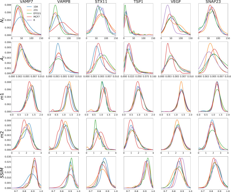
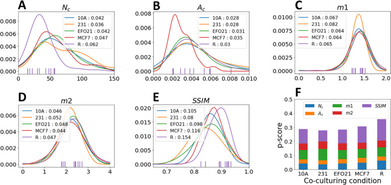
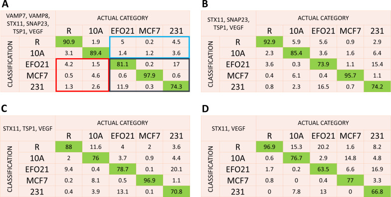
Results: A fast, streamlined and objective procedure for SRM platelet image acquisition, analysis and classification was developed to overcome these limitations. By stimulated emission depletion SRM we imaged nanoscale patterns of six different platelet proteins; four different SNAREs (soluble N-ethylmaleimide factor attachment protein receptors) mediating protein secretion by membrane fusion of storage granules, and two angiogenesis regulating proteins, representing cargo proteins within these granules coupled to tumor progression. By a streamlined procedure, we recorded about 100 SRM images of platelets, for each of these six proteins, and for five different categories of platelets; incubated with cancer cells (MCF-7, MDA-MB-231, EFO-21), non-cancer cells (MCF-10A), or no cells at all. From these images, structural similarity and protein cluster parameters were determined, and probability functions of these parameters were generated for the different platelet categories. By comparing these probability functions between the categories, we could identify nanoscale alterations in the protein distributions, allowing us to classify the platelets into their correct categories, if they were co-incubated with cancer cells, non-cancer cells, or no cells at all.

Conclusions: The fast, streamlined and objective acquisition and analysis procedure established in this work confirms the role of SNAREs and angiogenesis-regulating proteins in platelet-mediated cancer progression, provides additional fundamental knowledge on the interplay between tumor cells and platelets, and represent an important step towards using tumor-platelet interactions and redistribution of nanoscale protein patterns in platelets as a basis for cancer diagnostics.

Keywords: Cancer; Dictionary learning; Platelet; SNARE protein; STED; Super-resolution microscopy; Tumorigenesis.


Conflict of interest statement

The authors declare that they have no competing interests.

Figures

Fig. 1
Fig. 1
Representative STED images of platelets with different protein labels and subject to different co-culturing conditions. Platelets with different protein labels are imaged either under resting (R) condition, after co-culturing with cells from a non-tumor cell line MCF-10A (10A), or from different tumor cell lines MDA-MB-231 (231), EFO-21 (EFO21) and MCF-7 (MCF7). Scale bar: 1 µm
Fig. 2
Fig. 2
Overall workflow for the acquisition of platelet images, analysis of nanoscale protein distribution patterns and classification based on probabilities for image parameters measured. See text for more details
Fig. 3
Fig. 3
Normalized KDE probability functions for the different platelet image parameters, for each protein studied and under each of the different co-culturing conditions. The parameters are total number of clusters (Nc), mean area of cluster (Ac), first (m1) and second order moment (m2) of the radial distribution of clusters within a platelet, and the Structure Similarity Index Measure (SSIM), calculated by comparing the platelet image with its corresponding reconstructed image using dictionary learning (See text for more details). The x axis for each plot correspond to the respective parameter value (units: a.u for Nc and SSIM, µm2 for Ac and m2, µm for m1. The y axes show the normalized probability (a.u)
Fig. 4
Fig. 4
Example of p-score calculation (In this case based on images of platelets with VEGF staining). A set of 10 platelets from a certain (co-)culturing category are randomly selected (in this example from resting (R) platelets). For each of the 5 imaging parameters, the values determined for the 10 platelets are compared against the corresponding probability curves, in this example for VEGF, and for all the different platelet categories (AE). The probabilities of the 10 platelets to belong to a certain category are then added for each parameter (AE insets). F The combined p-score calculated for each co-culturing condition is obtained by adding the sum probabilities for all parameters for the given culturing category. The set of 10 platelets are finally classified into the category with the highest combined p-score, in this example into R
Fig. 5
Fig. 5
Classification probability matrices (in %) for individual proteins to classify a set of ten platelets of an actual category (columns) into a certain category (lines). The matrices are generated by randomly selecting 1000 sets of ten platelets from a known category and classifying them into one of the five categories. Probabilities enclosed in blue represent false negative classifications of tumor educated platelets
Fig. 6
Fig. 6
Classification matrices generated by combining the complementary classification strengths of different proteins. All possible combinations of 1 to 6 proteins were used for classification. The classification matrix for the best combination of A 6 proteins, B 4 proteins, C 3 proteins, and D 2 proteins. In A the probabilities enclosed in blue represent false negative classifications of tumor educated platelets (TEPs). Summing the probabilities enclosed in blue together, the average probability for false negative classification of TEPs, defined as the average probability to classify a platelet of category EFO21, MCF7 or 231 into the categories R or 10A is less than 6%. Similarly, the probabilities enclosed in red represent false positive classifications (with a probability < 8% when summed together), and those in black true positive classifications of TEPs (> 90% when summed together)

References

    1. Haemmerle M, Stone RL, Menter DG, Afshar-Kharghan V, Sood AK. The platelet lifeline to cancer: challenges and opportunities. Cancer Cell. 2018;33(6):965–983. doi: 10.1016/j.ccell.2018.03.002. - DOI - PMC - PubMed
    1. Tesfamariam B. Involvement of platelets in tumor cell metastasis. Pharmacol Therap. 2016;157(Supplement C):112–9. doi: 10.1016/j.pharmthera.2015.11.005. - DOI - PubMed
    1. Braun A, Anders HJ, Gudermann T, Mammadova-Bach E. Platelet-cancer interplay: molecular mechanisms and new therapeutic avenues. Front Oncol. 2021 doi: 10.3389/fonc.2021.665534. - DOI - PMC - PubMed
    1. Sabrkhany S, Kuijpers MJE, Egbrink M, Griffioen AW. Platelets as messengers of early-stage cancer. Cancer Metastasis Rev. 2021;40(2):563–573. doi: 10.1007/s10555-021-09956-4. - DOI - PMC - PubMed
    1. Meikle CKS, Kelly CA, Garg P, Wuescher LM, Ali RA, Worth RG. Cancer and thrombosis: the platelet perspective. Front Cell Dev Biol. 2017 doi: 10.3389/fcell.2016.00147. - DOI - PMC - PubMed

MeSH terms