

Vascular anomaly diagnosis by central venous catheter misplacement: a case report
- PMID: 35729662
- PMCID: PMC9215029
- DOI: 10.1186/s13256-022-03467-8
Vascular anomaly diagnosis by central venous catheter misplacement: a case report
Abstract
Background: Congenital heart diseases rarely have a primary manifestation in adulthood. They are a rare cause of pulmonary hypertension in adults.
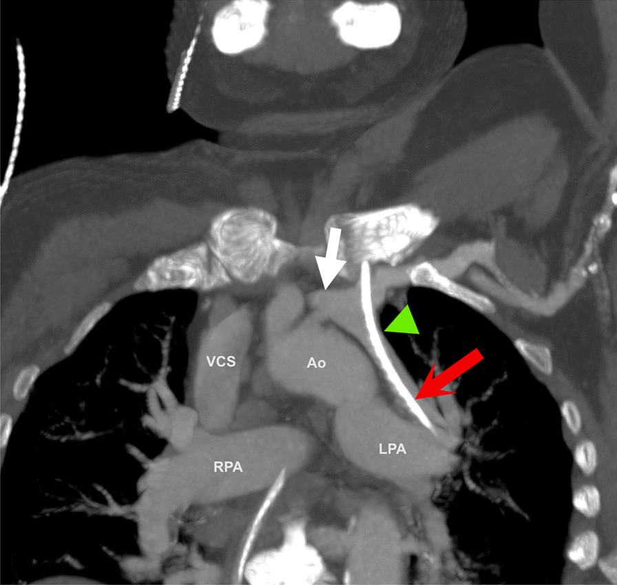
Case presentation: A 70-year-old woman of Eurasian descent underwent emergency surgery for bowel ischemia. Her history of mild pulmonary hypertension likely correlates with a peculiar diagnosis of an anatomic anomaly on the postoperative x-ray and computed tomography scan. The central venous catheter was misplaced. Initial management consisted of removal of the catheter. The diagnosis, partial anomalous pulmonary venous return, may pose a clinical therapeutic dilemma.
Conclusions: Partial anomalous pulmonary venous return is a potentially treatable cause of pulmonary hypertension. With the current trend toward more medical imaging, we expect this diagnosis to be made more often in the future.
Keywords: Cardiac vascular anatomy; Central venous catheter; Congenital heart disease; Pulmonary hypertension.
© 2022. The Author(s).
Conflict of interest statement
The authors declare that they have no competing interests.
Figures

References
-
- Gupta DL, Dwivedi DG. Entrapment of a guide wire: an incidental complication of central line catheter placement. Indian J Clin Anaesth. 2014;1:56–59.
Publication types
MeSH terms
LinkOut - more resources
Full Text Sources
Medical
