. 2022 Jul;15(7):e008997.
doi: 10.1161/CIRCHEARTFAILURE.121.008997. Epub 2022 Jun 22.

Estrogen Receptor-β Agonists Modulate T-Lymphocyte Activation and Ameliorate Left Ventricular Remodeling During Chronic Heart Failure

Affiliations

Estrogen Receptor-β Agonists Modulate T-Lymphocyte Activation and Ameliorate Left Ventricular Remodeling During Chronic Heart Failure

Rachel Rosenzweig et al. Circ Heart Fail. 2022 Jul.

Abstract

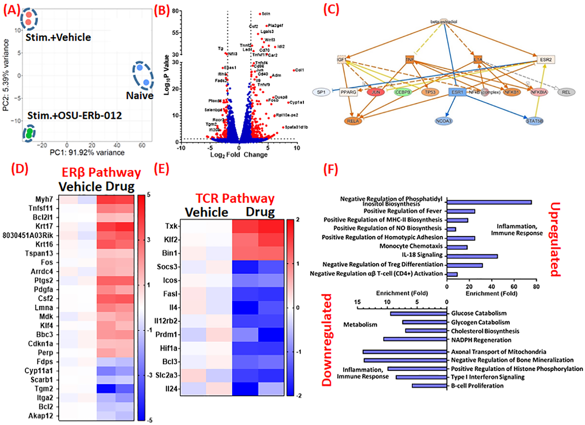
Background: CD4+ T cells temporally transition from protective to pathological during ischemic heart failure (HF; 8 weeks postmyocardial infarction). Cellular mechanisms mediating this shift are unknown.

Methods: RNA-sequencing of cardiac CD4+ T cells and flow cytometric analysis of immune cells was conducted.

Results: RNA-sequencing of CD4+ T cells from the failing hearts of male mice indicated activation of ER (estrogen receptor)-α signaling. Flow cytometric analysis showed that ERα in CD4+ T cells decreases significantly at 3-day postmyocardial infarction but increases during HF. To antagonize ERα, we tested a novel ERβ agonist (OSU-ERb-012) to inhibit T cells and blunt left ventricular remodeling. Proliferation assays showed that OSU-ERb-012 dose-dependently inhibited proliferation and proinflammatory cytokine expression in anti-CD3/CD28 stimulated splenic T cells isolated from both the sexes. For in vivo efficacy, 10- to 12-week-old male and ovariectomized female mice were randomized at 4 weeks postmyocardial infarction and treated with either vehicle or drug (60 mg/kg per day; oral). While vehicle-treated HF mice displayed progressive left ventricular dilatation with significantly increased end-systolic and end-diastolic volumes from 4 to 8 weeks postmyocardial infarction, treatment with OSU-ERb-012 significantly blunted these changes and stopped left ventricular remodeling in both the sexes. Reduction in tibia-normalized heart and left ventricular weights, cardiomyocyte hypertrophy and interstitial fibrosis further supported these results. Additionally, OSU-ERb-012 treatment selectively inhibited cardiac, splenic, and circulating CD4+ T cells without affecting other myeloid and lymphoid cells in the HF mice.

Conclusions: Our studies indicate that ERβ agonists and OSU-ERb-012, in particular, could be used as selective immunomodulatory drugs to inhibit CD4+ T cells during chronic HF.

Keywords: T-lymphocytes; heart failure; myocardial infarction; receptor, estrogen; ventricular remodeling.


Conflict of interest statement

Declarations of Interest: None

Figures

Figure 1:
Figure 1:
A) At 8 weeks post-myocardial infarction (MI), CD4+ T-cells from the failing hearts (150 cells) and mediastinal lymph-nodes (300 cells) were flow sorted and limited-cell RNA sequencing (lcRNA seq) was conducted to identify differential gene expression changes. IPA analysis identified ESR1 activation as one of the upregulated nodes in cardiac CD4+ T-cells and ESR1 dependent gene expression changes in the dataset are indicated. Upregulated genes are shown in red while downregulated genes are shown in green. Orange color represents ‘predicted activation’ whereas blue represents ‘predicted inhibition’. Lines in orange and blue represent ‘leads to activation or inhibition’ respectively, whereas yellow lines show ‘findings inconsistent with state of downstream molecule’ and grey lines show effects that could not be predicted. (B) Representative flow histograms showing ERα and ERβ expression in cardiac CD4+ T-cells at 3 days post-MI. Group quantitation for ERα (C) and ERβ (D) expression in cardiac CD4+ T-cells at 3 days and at 8 weeks post-MI. (E) Representative flow histograms showing ERβ expression in splenic (left) and cardiac (right) immune cells of myeloid and lymphoid lineages at 3 days post-MI. Group quantitation for mean fluorescence intensity (MFI) of ERβ in CD19+ B-cells, CD4+ T-cells, CD11b+Ly6G+ neutrophils and CD11b+Ly6GLy6C+ monocytes in the spleens (F) and the hearts (G) at 3 days (left) and at 8 weeks post-MI (right). Group quantitation for mean fluorescence intensity (MFI) of ERβ in CD4+ T-cells in the spleen, blood and the hearts at 3 days (H) and at 8 weeks post-MI (I). Data in C and D was analyzed using two-tailed unpaired students T-test whereas 1-way ANOVA with Tukey’s post-hoc test was used to analyze data in F, G, H and I.
Figure 2:
Figure 2:
(A) Representative flow cytometric histograms for Tag-it violet (TIV) labeled CD4+ T-cells either non-stimulated or stimulated with anti-CD3 and anti-CD28 antibodies in the absence and presence of different concentrations of OSU-ERb-012. Peak patterns from high to low fluorescence intensity in stimulated cells represent halving of dye concentration in the daughter cells with every successive cell division. Cell proliferation (%) in the presence of different concentrations of the drug is used to derive dose-response curve (lower panel, right). (B) Representative flow cytometric histograms for TIV labeled CD4+ T-cells either non-stimulated or stimulated with anti-CD3 and anti-CD28 antibodies and treated with either Estradiol (E2; 5 and 50 nM) or OSU-ERb-012 (5 μM) or both, and group quantitation for % cell proliferation (right). (C) Representative flow histograms showing TNFα expression in non-stimulated and stimulated CD4+ T-cells treated either with estradiol or OSU-ERb-012 or both. Group quantitation for the frequency of CD4+TNFα+ and CD4+IFNγ+ cells is shown in (D) and (E), respectively. Mean values from 3–4 separate experiments (conducted by isolating splenic CD4+ T-cells from 3–4 male mice) done in triplicate are reported. One way Anova with Tukey’s post-hoc test was used for the data analysis. *p represent significance with respect to non-stimulated cells whereas $p represent significance with respect to vehicle treated stimulated cells.
Figure 3:
Figure 3:
(A) Principal Component Analysis of RNA transcriptomes of naïve and stimulated (stim) CD4+ T-cells treated either with the vehicle (DMSO) or OSU-ERb-012 (5 μM). (B) Volcano plot showing several genes (marked as red) are either upregulated or downregulated by more than 2-fold in stimulated CD4+ T-cells treated with OSU-ERb-012 (5 μM) as compared to the vehicle treatment. Some of the representative genes that showed either very high LogP values or very high fold-changes are labeled. (C) Ingenuity Pathway analysis of RNA transcriptomes of stimulated CD4+ T-cells treated either with the vehicle control (DMSO) or OSU-ERb-012 (5 μM). Heat maps depicting genes in the Estrogen Receptor (ER) β (D) and T-cell receptor (TCR) (E) pathway that are either significantly up- or down-regulated in stimulated CD4+ T-cells upon treatment with OSU-ERb-012 (5 μM). (F) Pathways that are most affected in stimulated CD4+ T-cells upon treatment with OSU-ERb-012. N=2/gp.
Figure 4:
Figure 4:
(A) Schematic for the experimental plan to test the efficacy of OSU-ERb-012 during acute phase of MI and during chronic HF. Random: Randomization; Echo: echocardiography. (B) Body weight (g) of mice that either underwent sham-operation or myocardial infarction (MI) followed by treatment either with the vehicle or OSU-ERb-012 (60 mg/kg/day; gavage). For clarity of data, SD is shown only for HF group treated with vehicle and was comparable in all groups. N=4–7 in each group (C) Kaplan-Meier curve showing mortality rate in MI mice treated either with the vehicle or drug. N=6–10 in each group (D) Representative B-mode tracings depicting systole and diastole of failing hearts at 4 w (at the time of randomization) and 8 weeks post-MI after treatment either with the vehicle or the drug. (E) Group quantitation for the change in end-systolic and end-diastolic volumes (ESV and EV), and the ejection fraction (EF) from 4 to 8 weeks post-MI after treatment either with the vehicle or the drug. Unpaired students two-tailed T-test was used for analyzing data in (E).
Figure 5:
Figure 5:
(A) Gravimetric data for tibia normalized heart and left-ventricular (LV) weights of sham and heart failure (HF) mice treated either with the vehicle or OSU-ERb-012. N=9–12 in each group. (B) Representative images of LV sections stained with FITC conjugated Wheat-germ agglutinin to show cardiac hypertrophy. Boxed area in the upper panel is shown in the lower panel, and the group quantitation for cardiomyocyte area is shown in right. N=4 in each group. (C) Gene expression of cardiac hypertrophy markers in the remote-zone LV of HF mice treated either with the vehicle or OSU-ERb-012 from 4 to 8 weeks post-MI. N=4–8 in each group. (D) Representative images showing interstitial fibrosis in the LVs of HF mice treated either with the vehicle or the drug and their group quantitation. N=4–5 in each group. Two way Anova with Tukey’s post-hoc test was used for data analysis in (A), while two-tailed students T-test was used for (B), (C) and (D).
Figure 6:
Figure 6:
(A) Representative flow scatter plots depicting CD4+ and CD8+ T-cells in total CD45+ cells. HF: heart failure. (B) Levels of CD4+ Helper T-cells and its pro-inflammatory subsets viz CD4+TNFα+ cells and CD4+IFNγ+ (Th1), T-cells at 8 weeks post-MI in mice treated either with the vehicle or OSU-ERb-012 from 4 to 8 weeks post-MI. (C) Representative flow scatter plots for splenic CD4+ and CD8+ T-cells in CD45+ leukocytes, and (D) group quantitation for splenic CD4+ Helper T-cells (total cell numbers and frequency) at 8 weeks post-MI in mice treated either with the vehicle or OSU-ERb-012 from 4 to 8 weeks post-surgery. Unpaired Students 2-tailed T-test was used for the data analysis in (B) and (D).

References

    1. Bansal SS, Ismahil MA, Goel M, Patel B, Hamid T, Rokosh G and Prabhu SD. Activated T Lymphocytes are Essential Drivers of Pathological Remodeling in Ischemic Heart Failure. Circ Heart Fail. 2017;10:e003688. - PMC - PubMed
    1. Ngwenyama N, Kirabo A, Aronovitz M, Velazquez F, Carrillo-Salinas F, Salvador AM, Nevers T, Amarnath V, Tai A, Blanton RM et al. Isolevuglandin-Modified Cardiac Proteins Drive CD4+ T-Cell Activation in the Heart and Promote Cardiac Dysfunction. Circulation. 2021;143:1242–1255. - PMC - PubMed
    1. Carrillo-Salinas FJ, Ngwenyama N, Anastasiou M, Kaur K and Alcaide P. Heart Inflammation: Immune Cell Roles and Roads to the Heart. Am J Pathol. 2019;189:1482–1494. - PMC - PubMed
    1. Nehra S, Gumina RJ and Bansal SS. Immune cell Dilemma in Ischemic Cardiomyopathy: To Heal or Not to Heal. Curr Opin Physiol. 2021;19:39–46. - PMC - PubMed
    1. Ismahil MA, Hamid T, Bansal SS, Patel B, Kingery JR and Prabhu SD. Remodeling of the mononuclear phagocyte network underlies chronic inflammation and disease progression in heart failure: critical importance of the cardiosplenic axis. Circ Res. 2014;114:266–82. - PMC - PubMed

Publication types