. 2022 Jun 22;7(12):e158754.
doi: 10.1172/jci.insight.158754.

Blocking cell cycle progression through CDK4/6 protects against chronic kidney disease

Affiliations

Blocking cell cycle progression through CDK4/6 protects against chronic kidney disease

Yosuke Osaki et al. JCI Insight. .

Abstract

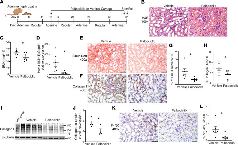
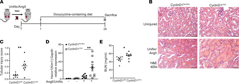
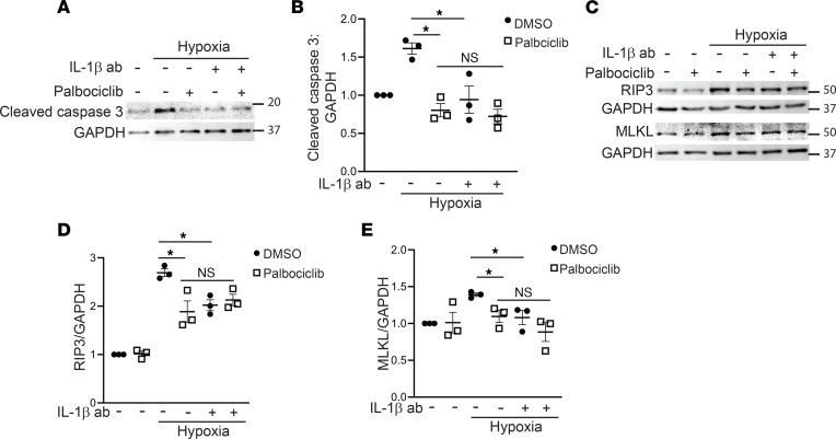
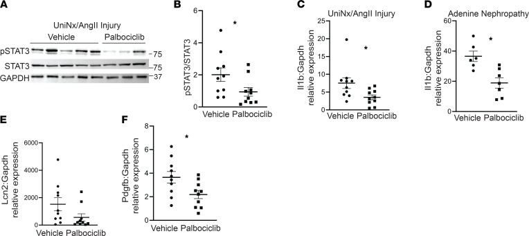
Acute and chronic kidney injuries induce increased cell cycle progression in renal tubules. While increased cell cycle progression promotes repair after acute injury, the role of ongoing tubular cell cycle progression in chronic kidney disease is unknown. Two weeks after initiation of chronic kidney disease, we blocked cell cycle progression at G1/S phase by using an FDA-approved, selective inhibitor of CDK4/6. Blocking CDK4/6 improved renal function and reduced tubular injury and fibrosis in 2 murine models of chronic kidney disease. However, selective deletion of cyclin D1, which complexes with CDK4/6 to promote cell cycle progression, paradoxically increased tubular injury. Expression quantitative trait loci (eQTLs) for CCND1 (cyclin D1) and the CDK4/6 inhibitor CDKN2B were associated with eGFR in genome-wide association studies. Consistent with the preclinical studies, reduced expression of CDKN2B correlated with lower eGFR values, and higher levels of CCND1 correlated with higher eGFR values. CDK4/6 inhibition promoted tubular cell survival, in part, through a STAT3/IL-1β pathway and was dependent upon on its effects on the cell cycle. Our data challenge the paradigm that tubular cell cycle progression is beneficial in the context of chronic kidney injury. Unlike the reparative role of cell cycle progression following acute kidney injury, these data suggest that blocking cell cycle progression by inhibiting CDK4/6, but not cyclin D1, protects against chronic kidney injury.

Keywords: Cell cycle; Fibrosis; Nephrology.


Conflict of interest statement

Conflict of interest: The authors have declared that no conflicts of interest exist.

Figures

Figure 1
Figure 1. Injured proximal tubules lacking TGF-β receptor have increased cell cycle progression.
(A) Transcript levels of genes related to cell cycle progression from RNA-Seq on serum-starved innermedullary collecting duct cells ± the TGF-β type II receptor (TβRII). (B) Gene expression of Cdkn2b (p15) and Cdkn2a (p16) were measured in conditionally immortalized PT cells using qPCR with Gapdh as a housekeeping gene. (C and D) Cell cycle of the PT cells (i.e., GFP+) from mice with TβRII intact (γGT-Cre;mT/mG) compared with those with TβRII selectively deleted in PT cells (γGT-Cre;mT/mG;Tgfbr2fl/fl) at 1 (C) or 4 weeks (D) after AA injury. (E and F) Actively cycling cells were detected and quantified using the R26Fucci2aR (Fucci) reporter with IHC using GFP antibody to detect Venus+ (S, G2, M) proximal tubule cells in mice with TβRII intact (γGT-Cre;Fucci) and TβRII selectively deleted in the proximal tubule (γGT-Cre;Fucci;Tgfbr2fl/fl) in both the AA and UniNx/AngII models. Statistical significance was determined by Benjamini-Hochberg correction for multiple testing in A and using 2-tailed Student’s t test for others. *P < 0.05 and **P < 0.01. Scale bar: 50 μm. PT, proximal tubule; AA, aristolochic acid; UniNx/AngII, uninephrectomy/angiotensin II).
Figure 2
Figure 2. Blocking CDK4/6 reduces tubular injury, fibrosis, and senescence after UniNx/AngII.
(A and B) Diagram of cell cycle progression at G1/S and injury schematic for UniNx/AngII. (C and D) UniNx/AngII-treated kidney lysates were blotted for retinoblastoma protein (Rb) phosphorylated at serine807/811, the target of CDK4/6. (E and F) H&E of kidneys injured by UniNx/AngII and treated with either gavage or palbociclib, a CDK4/6 inhibitor (E) and quantification of tubular injury (F). (G and H) Gene expression of KIM-1 from injured kidney cortices measured by qPCR and BUN from plasma at the time of sacrifice. (IL and NQ) Staining and quantification of Picrosirius red (I and J), collagen I (K and L), F4/80 (N and O), and TUNEL (P and Q). (M) Collagen I gene expression (Col1a1) measured from renal cortices using qPCR. (RT) Senescence assessed by β-galactosidase staining on frozen sections (R and S) and gene expression of p21 (Cdkn1a) on injured renal tissue (T). The percentage of β-galactosidase+ area was quantified on 400× fields (S). *P < 0.05,**P < 0.01, and ***P < 0.001 calculated by 2-tailed Student’s t test. Scale bar: 50 μm for 400× and 100 μm for 200×.
Figure 3
Figure 3. CDK4/6 inhibition ameliorates renal function and fibrosis after adenine nephropathy.
(A) Schematic for the adenine nephropathy model and palbociclib administration. (BD) Following adenine nephropathy, H&E of kidney cortices (B), BUN at time of sacrifice (C), and KIM-1 (Havcr1) gene expression in renal cortices by qPCR (D). (EJ) Picrosirius red and collagen I staining and quantification (EH), and collagen I immunoblots (collagen I MW of 130 kDa) on renal cortical lysates with α-tubulin as loading control and quantified (I and J). (K and L) F4/80 staining and quantification in renal cortices. *P < 0.05 calculated by 2-tailed Student’s t test, and scale bar: 50 μm.
Figure 4
Figure 4. Selective tubular deletion of cyclin D1 exacerbates UniNx/AngII-induced injury.
(A) Schematic of UniNx/AngII injury and recombination with doxycycline-containing diet. (B and C) H&E of renal cortices after UniNx/AngII injury and quantification of tubular injury. (D) Gene expression of KIM-1 (Havcr1) from injured renal cortices, normalized to Gapdh, was measured by qPCR. (E) BUN from plasma at the time of sacrifice. *P < 0.05, **P < 0.01 using 2-tailed Student’s t test. Scale bar: 50 μm.
Figure 5
Figure 5. Blocking CDK4/6 reduces tubule cell death.
(A and B) Immunoblots and quantification of cleaved caspase 3 from immortalized PT cells treated with siRNA to Cdnk2a (p16) or scramble and incubated with 30 μM AA for 3 days with GAPDH as a loading control. (CH) Primary PT cells were treated with 15 μM AA, angiotensin II 1 μM, or 1% O2 (hypoxia) for 7 days plus either palbociclib (2 μM) or equal volume DMSO (diluent) and immunoblotted for cleaved caspase 3 with quantification of 3 separate experiments. (I and J) Immortalized PT cells were transfected with siRNA to Rb or scramble, treated with 30 μM AA for 3 days, and immunoblotted for cleaved caspase 3 or GAPDH as loading control. *P < 0.05 using the 2-tailed Student’s t test. AA, aristolochic acid; Rb, retinoblastoma.
Figure 6
Figure 6. Palbociclib reduces STAT3 activation and downstream targets.
(AD) Primary PT cells were treated with hypoxia (1% O2) or 1 μM angiotensin II (AngII) ± 2 μM palbociclib for 7 days and immunoblotted for phosphorylated STAT3 (pSTAT3), total STAT3, or GAPDH for loading control. (E and F) Gene expression of IL-1β (Il1b), Cxcl2, and Cxcl5 from primary PT cells treated with hypoxia for 2 days or AngII for 7 days was measured by qPCR with Gapdh as a housekeeping gene. (G) Conditionally immortalized proximal tubule cells with and without TβRII were treated with the indicated concentrations of aristolochic acid (AA) for 4 days, and then cDNA was generated for IL-1β (Il1b) gene expression by qPCR. *P < 0.05, **P < 0.01, and ***P < 0.001 using the 2-tailed Student’s t test.
Figure 7
Figure 7. Palbociclib reduces tubular apoptosis and necroptosis, in part, through IL-1β.
(AE) Primary PT cells were treated with 1% O2 (hypoxia) for 7 days plus IL-1β blocking antibody (100 ng/mL), palbociclib (2 μM), or both, and lysates blotted for cleaved caspase 3 (A and B), or RIP3 and MLKL (necroptosis) and quantified with GAPDH as a loading control (CE). *P < 0.05 using ordinary 1-way ANOVA with GraphPad Prism. RIP3, receptor interacting-protein 3; MLKL, mixed lineage kinase domain-like protein.
Figure 8
Figure 8. Palbociclib reduces STAT3 activation and IL-1β expression in CKD injury models.
(A and B) Cortical tissue from mice injured by UniNx/AngII was immunoblotted for pSTAT3, total STAT3, and GAPDH for loading control and quantified. (C and D) Gene expression for IL-1β (Il1b) was measured in renal cortices of UniNx/AngII-injured and adenine nephropathy-treated mice with qPCR using Gapdh as a housekeeping gene. (E and F) NGAL (Lcn2) and PDGF-β (Pdgfb) were measured in renal cortices of UniNx/AngII injured mice. *P < 0.05 using the 2-tailed Student’s t test. NGAL, neutrophil gelatinase-associated lipocalin; PDGF-β, platelet-derived growth factor β.

Similar articles

Cited by

References

    1. Grgic I, et al. Targeted proximal tubule injury triggers interstitial fibrosis and glomerulosclerosis. Kidney Int. 2012;82(2):172–183. doi: 10.1038/ki.2012.20. - DOI - PMC - PubMed
    1. Takaori K, et al. Severity and frequency of proximal tubule injury determines renal prognosis. J Am Soc Nephrol. 2016;27(8):2393–2406. doi: 10.1681/ASN.2015060647. - DOI - PMC - PubMed
    1. Gewin L, et al. Progression of chronic kidney disease: too much cellular talk causes damage. Kidney Int. 2017;91(3):552–560. doi: 10.1016/j.kint.2016.08.025. - DOI - PMC - PubMed
    1. Ishibe S, Cantley LG. Epithelial-mesenchymal-epithelial cycling in kidney repair. Curr Opin Nephrol Hypertens. 2008;17(4):379–385. doi: 10.1097/MNH.0b013e3283046507. - DOI - PubMed
    1. Nlandu-Khodo S, et al. Blocking TGF- β and β-catenin epithelial crosstalk exacerbates CKD. J Am Soc Nephrol. 2017;28(12):3490–3503. doi: 10.1681/ASN.2016121351. - DOI - PMC - PubMed

Publication types