. 2022 Aug 31;10(4):e0132422.
doi: 10.1128/spectrum.01324-22. Epub 2022 Jun 22.

Mycobiome Dysbiosis in Women with Intrauterine Adhesions

Affiliations

Mycobiome Dysbiosis in Women with Intrauterine Adhesions

Ning-Ning Liu et al. Microbiol Spectr. .

Abstract

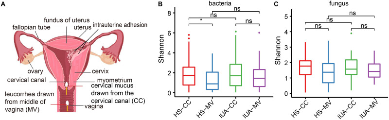
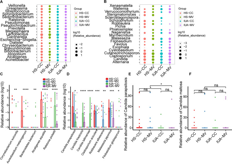
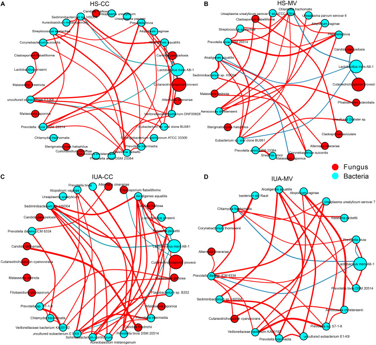
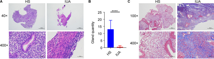
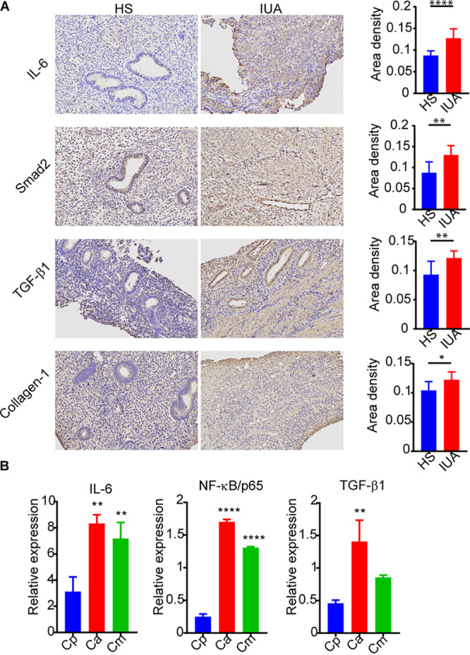
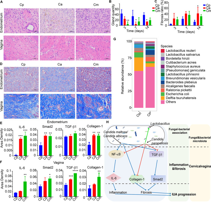
The vaginal microbiota dysbiosis is closely associated with the development of reproductive diseases. However, the contribution of mycobiome to intrauterine adhesion (IUA) disease remains unknown. Harnessing 16S and ITS2 rDNA sequencing analysis, we investigate both bacterial and fungal microbiota compositions across 174 samples taken from both cervical canal (CC) and middle vagina (MV) sites of IUA patients. Overall, there is no significant difference in microbial diversity between healthy subjects (HS) and IUA patients. However, we observe the IUA-specific bacterial alterations such as increased Dialister and decreased Bifidobacterium and enriched fungal genera like increased Filobasidium and Exophiala. Moreover, site-specific fungal-bacterial correlation networks are discovered in both CC and MV samples of IUA patients. Mechanistic investigation shows that Candida parapsilosis, other than Candida albicans and Candida maltosa, prevents the exacerbation of inflammatory activities and fibrosis, and modulates bacterial microbiota during IUA progression in a rat model of IUA. Our study thus highlights the importance of mycobiota in IUA progression, which may facilitate the development of therapeutic target for IUA prevention. IMPORTANCE Intrauterine adhesion (IUA) often leads to hypomenorrhea, amenorrhea, repeat miscarriages, and infertility. It has been prevalent over the last few decades in up to 13% of women who experience pregnancy termination during the first trimester, and 30% of women undergo dilation and curettage after a late, spontaneous abortion. However, the pathogenesis of IUA remains unclear. Despite reports of microbiota dysbiosis during IUA progression, there is little information on the effect of fungal microbiota on the development of IUA. This study not only enhances our understanding of the mycobiome in IUA patients but also provides potential intervention strategies for prevention of IUA by targeting mycobiome.

Keywords: dysbiosis; fungal-bacterial correlation; intrauterine adhesions; mycobiome; reproductive tract.


Conflict of interest statement

The authors declare no conflict of interest.

Figures

FIG 1
FIG 1
Altered diversity of both bacterial and fungal microbiota. (A) Diagram of the female reproductive tract showing intrauterine adhesions and sampling sites within the cervical canal (CC) and middle vagina (MV) sites. Density plots showing the α-diversity of bacteria (B) and fungi (C), presented as estimated by the Shannon index. HS, healthy subjects; IUA, intrauterine adhesion. *, P < 0.05; **, P < 0.01; ns, not significant. P values are determined by Kruskal-Wallis test.
FIG 2
FIG 2
Global composition of bacterial microbiota at the genus (A) and species (C) levels. Global composition of fungal microbiota at the genus (B) and species (D) levels. Relative abundance of Candida parapsilosis (E) and Candida maltosa (F) in the lower female reproductive tract (CC and MV) of IUA patients and HS. Average relative abundance is based on circle size. For A to E, IUA CC group, n = 43; IUA MV group, n = 43; HS CC group, n = 44; and HS MV group, n = 44. *, P < 0.05; **, P < 0.01; ***, P < 0.001; ****, P < 0.0001; ns, not significant. P values are determined by Kruskal-Wallis test.
FIG 3
FIG 3
Imbalanced fungal-bacterial correlation networks in both HS (A and B) and IUA patients (C and D) by CytoSCAPE. Each node represents a bacterial or fungal phylum. Node size represents the number of direct edges it has. Edge color indicates the magnitude of the distance correlation (red indicates positive correlation and green indicates negative correlation as determined by the Spearman test). Only the top 30 species in relative abundance with significant correlations (absolute value of correlation coefficient >0.4 and Benjamini-Hochberg adjusted P value < 0.05) are displayed.
FIG 4
FIG 4
H&E and Masson staining of endometrial tissue from IUA patients. (A) hematoxylin and eosin (H&E) staining of endometrial tissues in HS (left) and IUA patients (right) shown at ×40 and ×400 magnifications. (B) Bar chart illustrating differences in gland quantity for endometrial tissue from IUA patients (n = 10) and HS (n = 10). (C) Masson staining of endometrial tissue in HS (left) and IUA patients (right). In A to C: ****, P < 0.0001. P values are determined by two-tailed Student's t test (B).
FIG 5
FIG 5
Exacerbated inflammatory activities and fibrosis induced during IUA. (A) Immunohistochemistry assay of endometria tissues from IUA patients and healthy subjects. (B) Bar graphs showing inteluekin-6 (IL-6), NF-κB/p65, and transforming growth factor-β1 (TGF-β1) expression in primary endometrial cells stimulated by Candida albicans (Ca), C. parapsilosis (Cp), and C. maltose (Cm) as quantified by quantitative RT-PCR. In A and B: *, P < 0.05; **, P < 0.01; ****, P < 0.0001. The P values are determined by two-tailed Student’s t test (A) and one-way ANOVA and Dunnett post hoc tests (B).
FIG 6
FIG 6
Protective benefits of C. parapsilosis against IUA progression in rat models. (A) H&E staining of rat endometrial and vaginal tissues containing each of three Candida species. (B) Bar graphs showing representative gland quantity in endometria of rats from each Candida group. (C) Masson staining of rat endometrial and vaginal tissues exposed to each Candida species examined. (D) Bar graphs for representative fibrosis area ratios of endometria in rats for each Candida group over three time points. (E and F) Bar graphs showing immunohistochemistry results of Smad2, IL-6, and TGF-β1 in endometrial tissue (E) and vaginal tissue (F). (G) Global dysbiosis of bacterial microbiota composition at the species level for both control (left) and Cp (right) groups after 14 days (n = 6). (H) Diagram showing how mycobiome dysbiosis, especially from Candida spp, contributes to IUA progression through regulation of inflammation and fibrosis, and through change of bacterial microbiota. In all panels: *, P < 0.05; **, P < 0.01; ***, P < 0.001; ****, P < 0.0001. The P values are determined by one-way ANOVA and Dunnett post hoc tests (B, D, and E). Cp, Cp-treated group (n = 6); Ca, Ca-treated group (n = 6); Cm, Cm-treated group (n = 6).

Similar articles

Cited by

References

    1. Deans R, Abbott J. 2010. Review of intrauterine adhesions. J Minim Invasive Gynecol 17:555–569. doi:10.1016/j.jmig.2010.04.016. - DOI - PubMed
    1. Doroftei B, Dabuleanu AM, Ilie OD, Maftei R, Anton E, Simionescu G, Matei T, Armeanu T. 2020. Mini-review of the new therapeutic possibilities in asherman syndrome–where are we after one hundred and twenty-six years? Diagnostics 10:706. doi:10.3390/diagnostics10090706. - DOI - PMC - PubMed
    1. Dreisler E, Kjer JJ. 2019. Asherman’s syndrome: current perspectives on diagnosis and management. Int J Womens Health 11:191–198. doi:10.2147/IJWH.S165474. - DOI - PMC - PubMed
    1. Wang YQ, Song XH, Wu SL, Huang YZ, Yan L, Li CZ. 2020. Comparison of autocross-linked hyaluronic acid gel and intrauterine device for preventing intrauterine adhesions in infertile patients: a randomized clinical trial. Gynecol Minim Invasive Ther 9:74–80. doi:10.4103/GMIT.GMIT_103_19. - DOI - PMC - PubMed
    1. Yu D, Wong YM, Cheong Y, Xia E, Li TC. 2008. Asherman syndrome–one century later. Fertil Steril 89:759–779. doi:10.1016/j.fertnstert.2008.02.096. - DOI - PubMed

Publication types