. 2022 Jul;28(7):1384-1392.
doi: 10.3201/eid2807.212614.

Nipah Virus Detection at Bat Roosts after Spillover Events, Bangladesh, 2012-2019

Nipah Virus Detection at Bat Roosts after Spillover Events, Bangladesh, 2012-2019

Clifton D McKee et al. Emerg Infect Dis. 2022 Jul.

Abstract

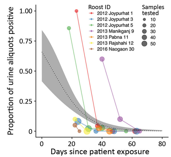
Knowledge of the dynamics and genetic diversity of Nipah virus circulating in bats and at the human-animal interface is limited by current sampling efforts, which produce few detections of viral RNA. We report a series of investigations at Pteropus medius bat roosts identified near the locations of human Nipah cases in Bangladesh during 2012-2019. Pooled bat urine was collected from 23 roosts; 7 roosts (30%) had >1 sample in which Nipah RNA was detected from the first visit. In subsequent visits to these 7 roosts, RNA was detected in bat urine up to 52 days after the presumed exposure of the human case-patient, although the probability of detection declined rapidly with time. These results suggest that rapidly deployed investigations of Nipah virus shedding from bat roosts near human cases could increase the success of viral sequencing compared with background surveillance and could enhance understanding of Nipah virus ecology and evolution.

Keywords: Bangladesh; Chiroptera; Henipavirus; Nipah virus; Pteropodidae; cross-species transmission; spillover; surveillance; viruses; zoonoses; zoonotic pathogens.


Figures

Figure 1
Figure 1
Locations of human Nipah cases (n = 21) and Pteropus medius bat roosts (n = 30) investigated in Bangladesh, 2012–2019. Roosts with urine aliquots that tested positive for Nipah virus RNA at the first sampling visit are indicated with triangles. Points have been jittered a small amount to increase visibility. Districts with human Nipah virus cases, identified bat roosts, or Nipah surveillance hospitals are labeled.
Figure 2
Figure 2
Descriptive variables for 23 Pteropus medius bat roosts sampled near confirmed human Nipah virus cases, Bangladesh, 2012–2019. Open circles show the values associated with the first human case associated with each roost; gray circles indicate means for each variable and positivity status (0 or 1). Vertical lines within boxes indicate medians; box left and right edges indicate the 25th and 75th percentiles; error bars indicate +1.5 times the interquartile range.
Figure 3
Figure 3
Results of screening of Pteropus medius bat roost urine aliquots for Nipah virus RNA, Bangladesh, 2012–2019. For each roost, the proportion of urine aliquots out of the total tested (indicated by the size of the circles) is aligned along a time axis of days since the first associated case-patient was exposed to Nipah virus in date palm sap. Time since patient exposure was either reported during the investigation or back-calculated as 7 days before reported symptom onset.

References

    1. Chadha MS, Comer JA, Lowe L, Rota PA, Rollin PE, Bellini WJ, et al. Nipah virus-associated encephalitis outbreak, Siliguri, India. Emerg Infect Dis. 2006;12:235–40. 10.3201/eid1202.051247 - DOI - PMC - PubMed
    1. Chua KB, Bellini WJ, Rota PA, Harcourt BH, Tamin A, Lam SK, et al. Nipah virus: a recently emergent deadly paramyxovirus. Science. 2000;288:1432–5. 10.1126/science.288.5470.1432 - DOI - PubMed
    1. Hsu VP, Hossain MJ, Parashar UD, Ali MM, Ksiazek TG, Kuzmin I, et al. Nipah virus encephalitis reemergence, Bangladesh. Emerg Infect Dis. 2004;10:2082–7. 10.3201/eid1012.040701 - DOI - PMC - PubMed
    1. Ching PKG, de los Reyes VC, Sucaldito MN, Tayag E, Columna-Vingno AB, Malbas FF Jr, et al. Outbreak of henipavirus infection, Philippines, 2014. Emerg Infect Dis. 2015;21:328–31. 10.3201/eid2102.141433 - DOI - PMC - PubMed
    1. Kessler MK, Becker DJ, Peel AJ, Justice NV, Lunn T, Crowley DE, et al. Changing resource landscapes and spillover of henipaviruses. Ann N Y Acad Sci. 2018;1429:78–99. 10.1111/nyas.13910 - DOI - PMC - PubMed

Publication types

LinkOut - more resources