. 2023 Jan;478(1):103-119.
doi: 10.1007/s11010-022-04487-0. Epub 2022 Jun 22.

Simulation of COVID-19 symptoms in a genetically engineered mouse model: implications for the long haulers

Affiliations

Simulation of COVID-19 symptoms in a genetically engineered mouse model: implications for the long haulers

Mahavir Singh et al. Mol Cell Biochem. 2023 Jan.

Abstract

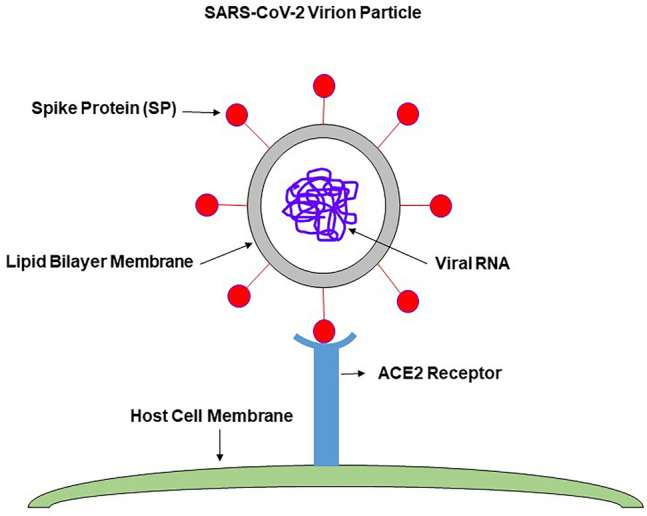
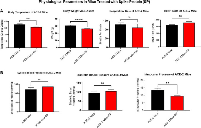
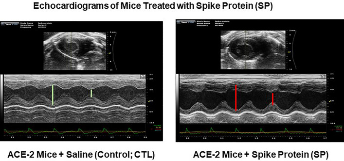
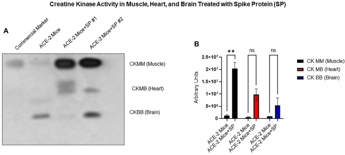
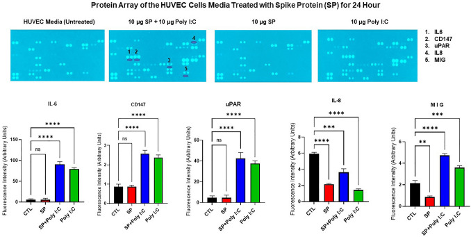
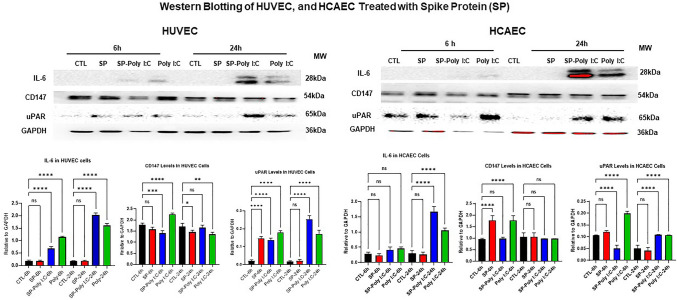
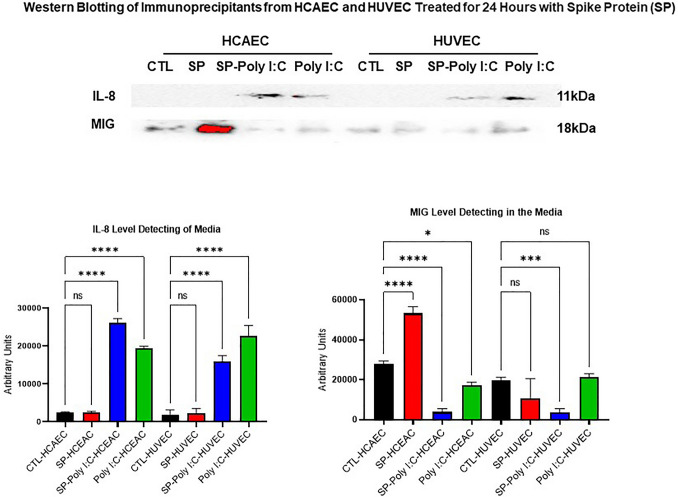
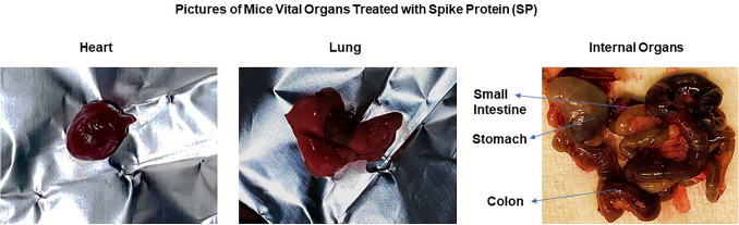
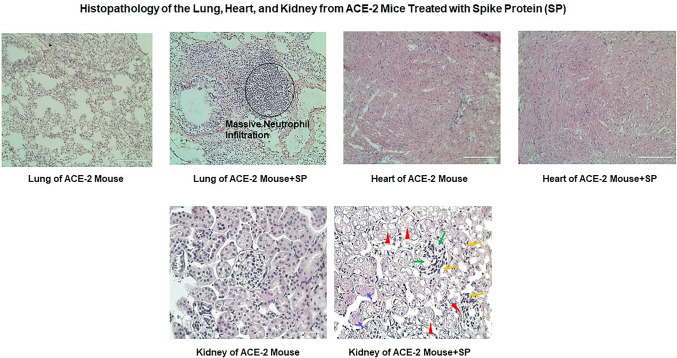
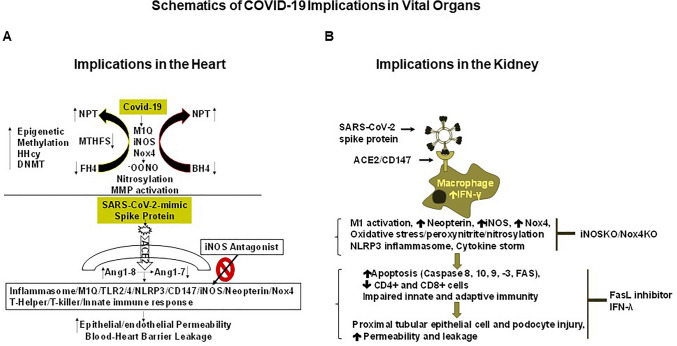
The ongoing pandemic (also known as coronavirus disease-19; COVID-19) by a constantly emerging viral agent commonly referred as the severe acute respiratory syndrome corona virus 2 or SARS-CoV-2 has revealed unique pathological findings from infected human beings, and the postmortem observations. The list of disease symptoms, and postmortem observations is too long to mention; however, SARS-CoV-2 has brought with it a whole new clinical syndrome in "long haulers" including dyspnea, chest pain, tachycardia, brain fog, exercise intolerance, and extreme fatigue. We opine that further improvement in delivering effective treatment, and preventive strategies would be benefited from validated animal disease models. In this context, we designed a study, and show that a genetically engineered mouse expressing the human angiotensin converting enzyme 2; ACE-2 (the receptor used by SARS-CoV-2 agent to enter host cells) represents an excellent investigative resource in simulating important clinical features of the COVID-19. The ACE-2 mouse model (which is susceptible to SARS-CoV-2) when administered with a recombinant SARS-CoV-2 spike protein (SP) intranasally exhibited a profound cytokine storm capable of altering the physiological parameters including significant changes in cardiac function along with multi-organ damage that was further confirmed via histological findings. More importantly, visceral organs from SP treated mice revealed thrombotic blood clots as seen during postmortem examination. Thus, the ACE-2 engineered mouse appears to be a suitable model for studying intimate viral pathogenesis thus paving the way for identification, and characterization of appropriate prophylactics as well as therapeutics for COVID-19 management.

Keywords: Clinical symptoms; Disease management; Humanized mouse; Multi-organ damage; SARS-CoV-2 spike protein.


Conflict of interest statement

The authors declare that they have no conflict of interest, financial or otherwise.

Figures

Fig. 1
Fig. 1
A schematic depicting the severe acute respiratory syndrome-coronavirus 2 (SARS-CoV-2) virion and binding of its spike protein (SP) with the host cell receptor. The SARS-CoV-2 ‘SP’ mediates binding of the virion with its receptor angiotensin converting enzyme 2 (ACE-2), and promotes fusion between the virion and host cell membrane thus allowing the virion entry into host cell. The viral ribonucleic acid (RNA) is a single stranded, and non-segmented that is ~ 30 kilobase in size is enclosed inside a protein coat known as the capsid. It is the capsid that is coated with ‘SP’ protein which has two subunits known as S1 and S2. The S2 subunit recognizes ACE-2 receptor on host cell membrane while S1 subunit helps mediate viral fusion with the cell
Fig. 2
Fig. 2
A Measurement of the body temperature, weight, respiration, heart rate, blood pressure (systolic, and diastolic), and intraocular pressure (IOP). The human angiotensin converting enzyme 2 transgenic mice (B6.Cg-Tg(K18-ACE2)2Prlmn/J, Genotype: Hemizygous genotype, Hemizygous for Tg(K18- ACE2)2Prlmn; in short, ACE-2 mice) were treated or untreated with the SARS-CoV-2 spike protein (SP). A Daily body temperature, and the body weight of the ACE-2 mice administered with SP via the nasal route were compared to the control mice (without SP). Unpaired t-tests were performed, *p < 0.01, **p < 0.001 ***p < 0.001, ****p < 0.0001, respectively, n = 3–5 mice/group. The respiration rate, and heart rate of ACE-2 mice (measured while doing the echocardiography) were also recorded. Unpaired t-test were performed, p < 0.2056, p < 0.2002, respectively, n = 4. B Blood pressure was measured by Coda non-invasive instrument. Unpaired t-tests were performed, p < 0.3890, n = 3–5 mice/group. Systolic, and diastolic pressure were measured, and the unpaired t-test were performed, p < 0.4696, n = 3–5 mice/group. Similarly, IOP was also measured by iCareLab tonometer, and unpaired t-test was performed, p < 0.0023, n = 3–5 mice/group. The body temperature is significantly decreased in mice treated with SP (35.50 ± 1.01 vs 31.39 ± 0.52), the body weight of those treated with SP were found to be significantly less (30.68 ± 0.73 vs 26.27 ± 0.42); however, the respiration rate (130.00 ± 7.4 vs 112.40 ± 18.7 breaths per minute), heart rate (321.70 ± 8.9 vs 358.00 ± 16.7 beats per minute), systolic (122.00 ± 8.3 vs 136.90 ± 8.56 mmHg), and diastolic blood pressure (93.33 ± 6.0 vs 105.00 ± 8.26 mmHg) were not significantly different. Interestingly, IOP was significantly decreased in mice treated with SP (13.43 ± 1.2 vs 9.500 ± 0.43)
Fig. 3
Fig. 3
Echocardiography of the SP treated ACE-2 mice versus untreated ACE-2 mice groups. Representative M-Mode image of parasternal long-axis view images from each group are presented indicating diastolic (longer) and systolic (shorter) chamber lengths in the ACE-2 mice treated with SP in comparison to the untreated control ACE-2 mice. The contraction and relaxation of the myocardium are attenuated in SP treated mice in comparison to the untreated control ACE-2 mice, n = 3–5 mice/group
Fig. 4
Fig. 4
Creatine phosphokinase (CK) levels in SP treated ACE-2) mice versus untreated ACE-2 mice. SP treated (SP #1 and SP #2) were higher compared to the control mice. Differences in CK levels (A) is due to multi-organ damage such as skeletal muscle (CK-MM), heart (CK-MB), and the brain (CK-BB) (B). The binding of SP to ACE-2 receptor causes multi-organ damage. Two-Way ANOVA with multiple comparisons, **p < 0.009, n = 3–5 mice/group
Fig. 5
Fig. 5
The proteome profiler with antibodies arrays reveals induction of cytokines in primary human umbilical vein endothelial cells (HUVEC). The cells were treated either with sterile phosphate buffered saline (PBS/saline) alone or SP; or with 10 mg of Poly I:C, or both SP and Poly I:C. The quantitation and comparison of SP induced CD147, IL-6 and IL-8, MIG, and uPAR are shown in comparison with respective controls. Depicted are the fluorescence intensity of different proteins measured, n = 3–5 petri dish/group
Fig. 6
Fig. 6
Western blot analyses of the key target proteins. Supernatants from human primary umbilical vein endothelial cells (HUVEC) at 6- and 24-h post treatment using the control (CTL), SP (spike protein), SP-Poly (spike protein and poly I:C), and Poly (poly I:C). The primary antibodies used were Interleukin-6 (IL-6), CD147 (EMPERIN), and uPAR and protein bands were normalized with GAPDH. The expression levels of each protein were also quantified as shown by the bar charts, n = 3–5 petri dish/group, ns not significant, *p < 0.01, ****p < 0.0001. Similarly, supernatants from human primary coronary artery endothelial cells (HCAEC) at 6- and 24-h post treatment were performed using the control (CTL), SP (spike protein), SP-Poly (spike protein and poly I:C), and Poly (poly I:C). The primary antibodies used were Interleukin-6 (IL-6), CD147 (EMPERIN), and uPAR, and the protein bands were normalized with GAPDH. The expression levels of each protein were also quantified as shown by the bar charts. n = 3–5 petri dish/group, ns not significant, *p < 0.01, ****p < 0.0001
Fig. 7
Fig. 7
Western blot analysis of the key target proteins employing the immuno-precipitate. Immunoprecipitants from the human primary coronary artery endothelial cells (HCAEC) and human primary umbilical vein endothelial cells (HUVEC) post 24 h treatment were used from the samples: control (CTL), SP (spike protein), SP-Poly (spike protein and poly I:C), and Poly (poly I:C). The primary antibodies used were for Interleukin-8 (IL-8), and MIG (cxcl9). Expression levels of the proteins are shown in the bar charts after the bands were normalized with GAPDH, n = 3–5 petri dish/group, ns not significant, *p < 0.01, ****p < 0.0001
Fig. 8
Fig. 8
Representative pictures of the mice heart, lung, and internal visceral organs. Heart, and intestine show most likely the evidence of blood clots, and thrombi formation. The vital organs look very dark in color indeed, n = 3–5 mice/group
Fig. 9
Fig. 9
Hematoxylin and eosin (H&E) staining of the lung, heart, and kidney samples from the humanized ACE-2 (B6. Cp-Tg) and ACE2 (B6.Cp-Tg) + Spike protein. Black circle clearly depicts a cluster of infiltrated immune cell populations while heart Sect. (5 μm thickness) shows diffused inflammatory cells throughout the parenchyma, magnification × 20, scale bar—50 μm n = 3–5 mice/group. Kidney Sects. (5 μm thickness) showing representative image of control kidney and spike protein (SP) treated mice. Pictures depict loss of glomerular tuft and hyaline deposit (green arrows), desquamation of tubular epithelium and necrosis (red arrow heads), inflammatory cell infiltration (yellow arrows), and tubular necrosis (purple arrow). Magnification × 60, scale bar—50 μm. The control mice received saline/PBS, n = 3–5 mice/group
Fig. 10
Fig. 10
Schematics of plausible hypothesis regarding SARS-CoV-2 induced visceral organ damage. The binding of SARS-CoV-2 spike protein (SP) with ACE-2 receptor mimics SARS-CoV-2 infection, and causes the accumulation of Ang1-8, activation of inflammasome, and M1Q macrophages via the “TLR4/NLRP3/CD147/Nox4/iNOS/neopterin” axis in the heart. This cascade of events leads to endothelial blood-heart barrier (BHB) leakage; however, the iNOSKO/Nox4KO and iNOS antagonists may help mitigate the inflammasome/NLRP3/M1Q mediated endothelial BHB leakage (A), as reported earlier by Tyagi and Singh, Multi-organ damage by COVID-19: Congestive (cardio-pulmonary) heart failure, and blood-heart barrier leakage, Mol Cell Biochem. 2021;476 (4):1891–1895). Similarly, biding of the SARS-CoV-2 spike protein (SP) to ACE-2/CD147 on macrophages can cause M1Q activation by IFN-γ toward generating the neopterin, and thus stimulating the iNOS, Nox4, and NLRP3 inflammasome pathway in the kidney that in turn can trigger apoptosis which may lead to CD4+ and CD8+ cell lymphopenia. These alterations might inflict the proximal tubular epithelial cell/podocyte damage, and the resultant parenchymal leakage. In that case, the iNOSKO/Nox4KO, and Fas/FasL antagonists (Kp7-6)/IFN-λ treatment could help mitigate the cytokine storm, and T cell lymphopenia thus protecting the proximal tubular epithelial/podocyte function (B). M1; inflammatory macrophage (M1Q), iNOS; inducible of nitric oxide synthase, BH4; tetrahydrobiopterin, FH4; tetrahydrofolate

Similar articles

Cited by

References

    1. Morens DM, Breman JG, Calisher CH, Doherty PC, Hahn BH, Keusch GT, Kramer LD, LeDuc JW, Monath TP, Taubenberger JK. The origin of COVID-19 and why it matters. Am J Trop Med Hyg. 2020;103:955–959. doi: 10.4269/ajtmh.20-0849. - DOI - PMC - PubMed
    1. Umakanthan S, Sahu P, Ranade AV, Bukelo MM, Rao JS, Abrahao-Machado LF, Dahal S, Kumar H, Kv D. Origin, transmission, diagnosis and management of coronavirus disease 2019 (COVID-19) Postgrad Med J. 2020;96:753–758. doi: 10.1136/postgradmedj-2020-138234. - DOI - PMC - PubMed
    1. Huang C, Wang Y, Li X, Ren L, Zhao J, Hu Y, Zhang L, Fan G, Xu J, Gu X, Cheng Z, Yu T, Xia J, Wei Y, Wu W, Xie X, Yin W, Li H, Liu M, Xiao Y, Gao H, Guo L, Xie J, Wang G, Jiang R, Gao Z, Jin Q, Wang J, Cao B. Clinical features of patients infected with 2019 novel coronavirus in Wuhan, China. Lancet. 2020;395:497–506. doi: 10.1016/s0140-6736(20)30183-5. - DOI - PMC - PubMed
    1. Singh M, Bhat PP, Mishra BP, Singh RK. Biological transmissibility of buffalopox virus. J Appl Anim Res. 1996;9:79–88. doi: 10.1080/09712119.1996.9706107. - DOI
    1. Du L, He Y, Zhou Y, Liu S, Zheng BJ, Jiang S. The spike protein of SARS-CoV–a target for vaccine and therapeutic development. Nat Rev Microbiol. 2009;7:226–236. doi: 10.1038/nrmicro2090. - DOI - PMC - PubMed

Substances