

Genetically Modified Porcine-to-Human Cardiac Xenotransplantation
- PMID: 35731912
- PMCID: PMC10361070
- DOI: 10.1056/NEJMoa2201422
Genetically Modified Porcine-to-Human Cardiac Xenotransplantation
Abstract
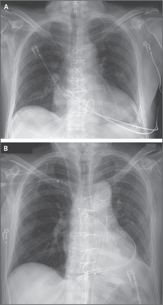
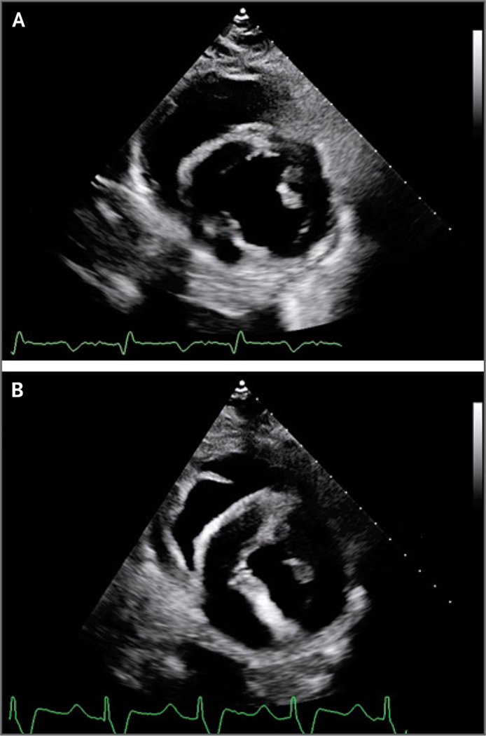
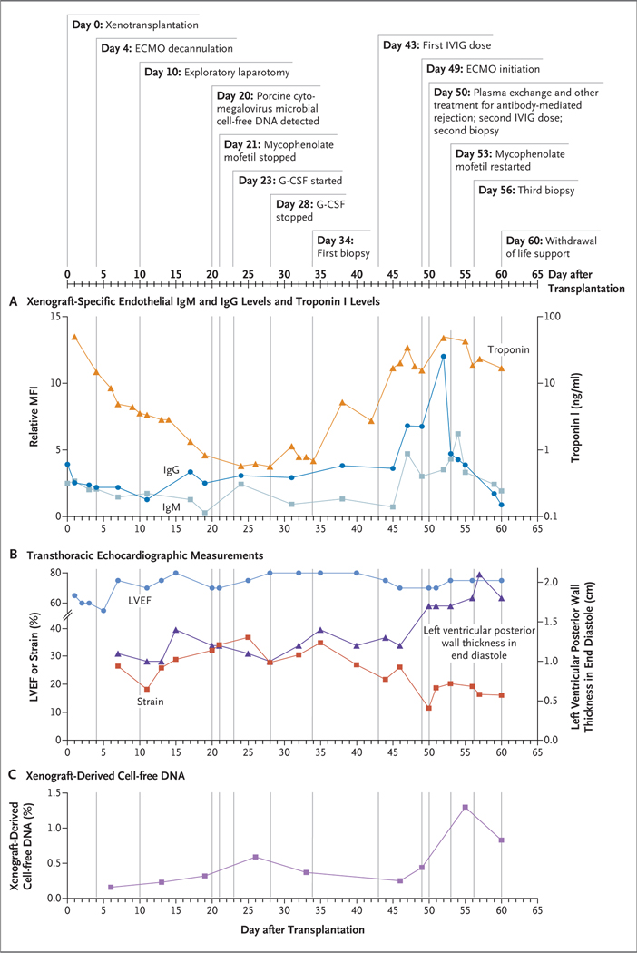
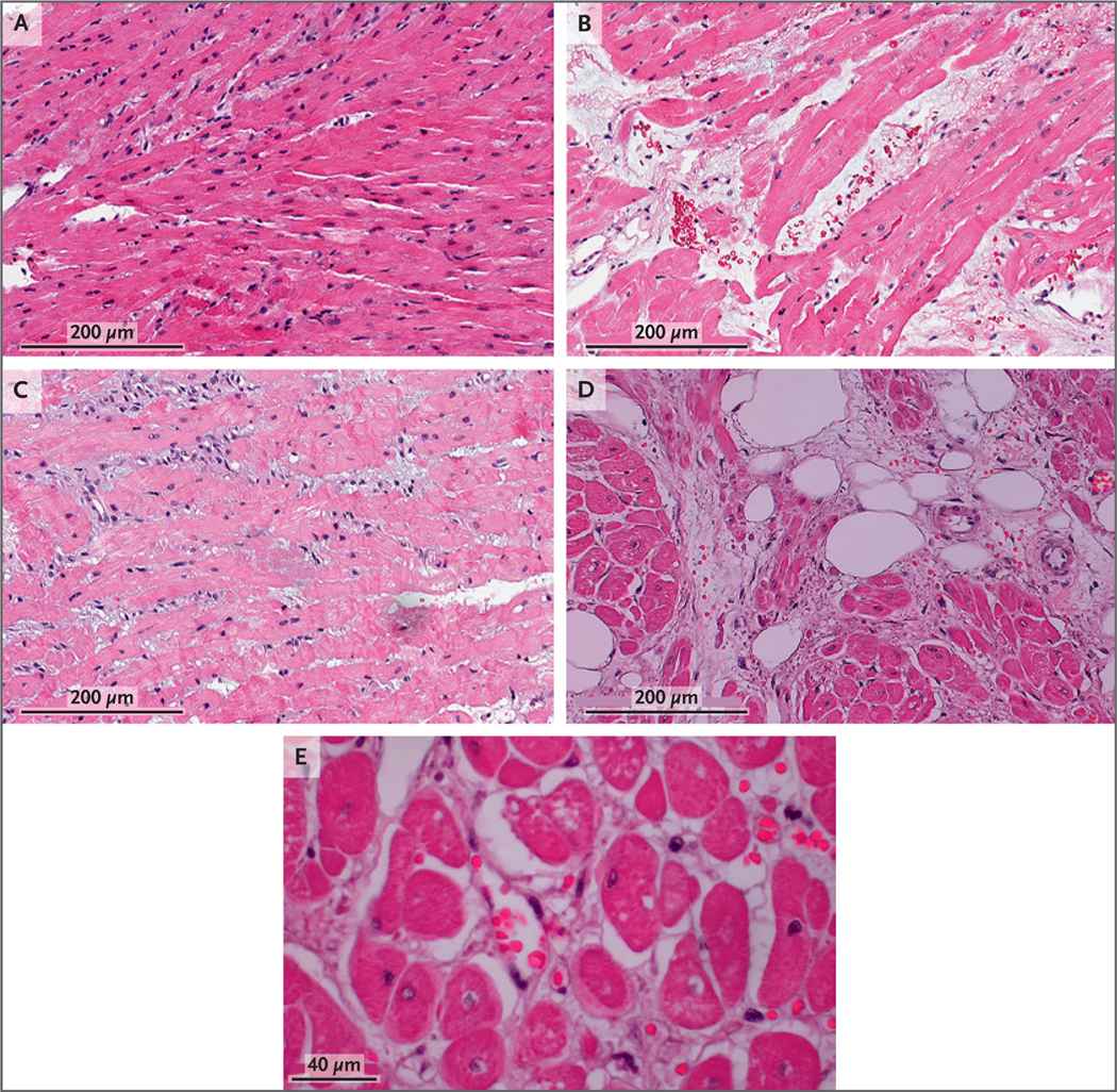
A 57-year-old man with nonischemic cardiomyopathy who was dependent on venoarterial extracorporeal membrane oxygenation (ECMO) and was not a candidate for standard therapeutics, including a traditional allograft, received a heart from a genetically modified pig source animal that had 10 individual gene edits. Immunosuppression was based on CD40 blockade. The patient was weaned from ECMO, and the xenograft functioned normally without apparent rejection. Sudden diastolic thickening and failure of the xenograft occurred on day 49 after transplantation, and life support was withdrawn on day 60. On autopsy, the xenograft was found to be edematous, having nearly doubled in weight. Histologic examination revealed scattered myocyte necrosis, interstitial edema, and red-cell extravasation, without evidence of microvascular thrombosis - findings that were not consistent with typical rejection. Studies are under way to identify the mechanisms responsible for these changes. (Funded by the University of Maryland Medical Center and School of Medicine.).
Copyright © 2022 Massachusetts Medical Society.
Figures
Comment in
-
Genetic Modification in Pig-to-Human Transplantation.N Engl J Med. 2022 Jul 7;387(1):79-82. doi: 10.1056/NEJMe2207422. Epub 2022 Jun 22. N Engl J Med. 2022. PMID: 35731913 No abstract available.
-
Genetically Modified Porcine-to-Human Cardiac Xenotransplantation.N Engl J Med. 2022 Oct 6;387(14):1337. doi: 10.1056/NEJMc2210401. N Engl J Med. 2022. PMID: 36198188 No abstract available.
-
Genetically Modified Porcine-to-Human Cardiac Xenotransplantation.N Engl J Med. 2022 Oct 6;387(14):1337-1338. doi: 10.1056/NEJMc2210401. N Engl J Med. 2022. PMID: 36198189 No abstract available.
References
-
- Eyestone W, Adams K, Ball S, et al. Gene-edited pigs for xenotransplantation. In: Cooper DKC, Byrne G, eds. Clinical xenotransplantation: pathways and progress in the transplantation of organs and tissues between species. Cham, Switzerland: Springer, 2020: 121–40.
-
- Hinrichs A, Riedel EO, Klymiuk N, et al. Growth hormone receptor knockout to reduce the size of donor pigs for preclinical xenotransplantation studies. Xenotransplantation 2021; 28: (2)e12664. - PubMed
-
- Diamond LE, Quinn CM, Martin MJ, Lawson J, Platt JL, Logan JS. A human CD46 transgenic pig model system for the study of discordant xenotransplantation. Transplantation 2001; 71: 132–42. - PubMed
Publication types
MeSH terms
Grants and funding
LinkOut - more resources
Full Text Sources
Other Literature Sources
Medical
Research Materials