. 2022 Jul 4;19(7):2287-2298.
doi: 10.1021/acs.molpharmaceut.2c00121. Epub 2022 Jun 22.

Binding of the Dual-Action Anti-Parkinsonian Drug AG-0029 to Dopamine D2 and Histamine H3 Receptors: A PET Study in Healthy Rats

Affiliations

Binding of the Dual-Action Anti-Parkinsonian Drug AG-0029 to Dopamine D2 and Histamine H3 Receptors: A PET Study in Healthy Rats

Nafiseh Ghazanfari et al. Mol Pharm. .

Abstract

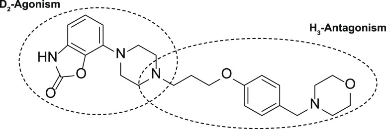
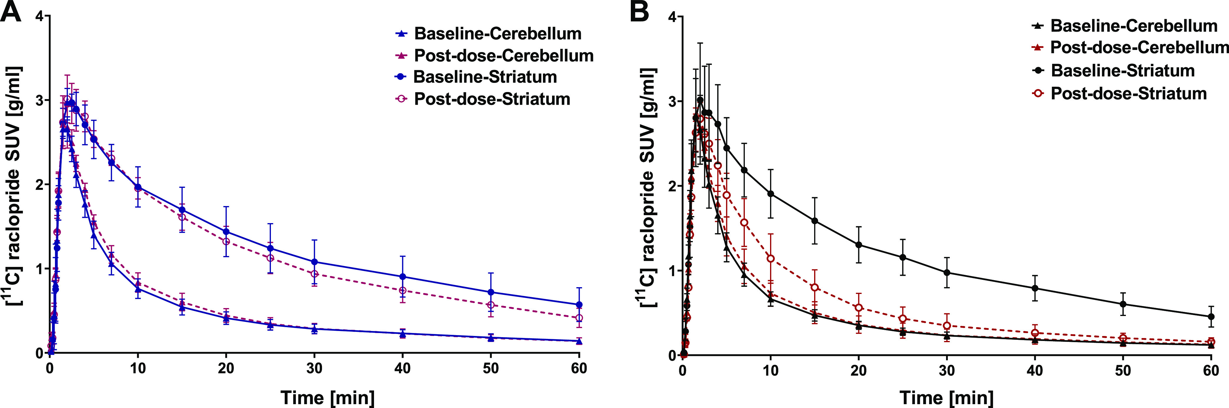
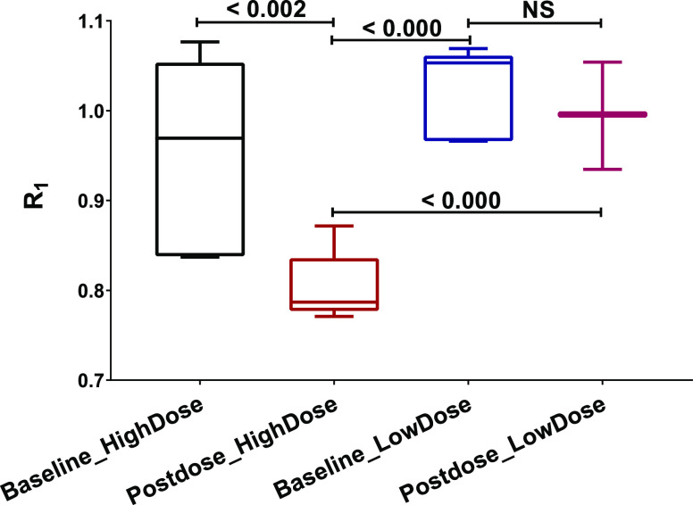
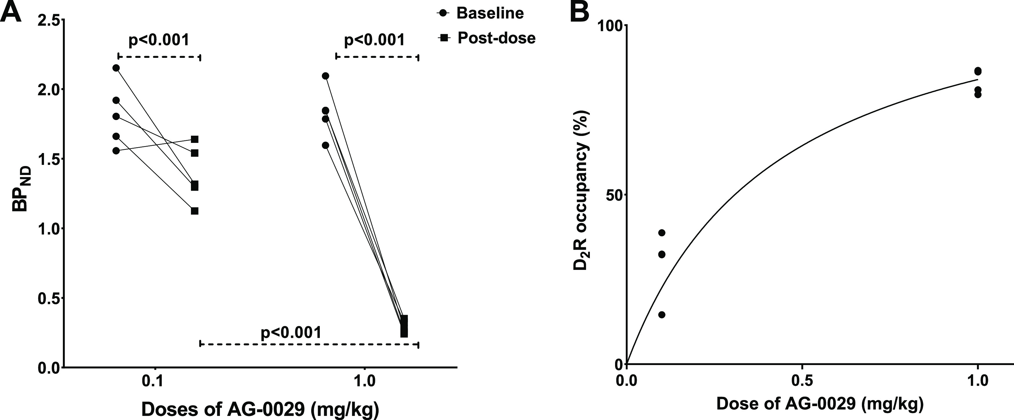
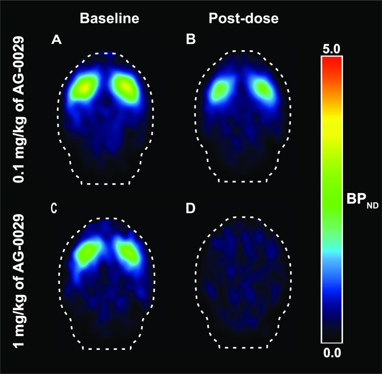
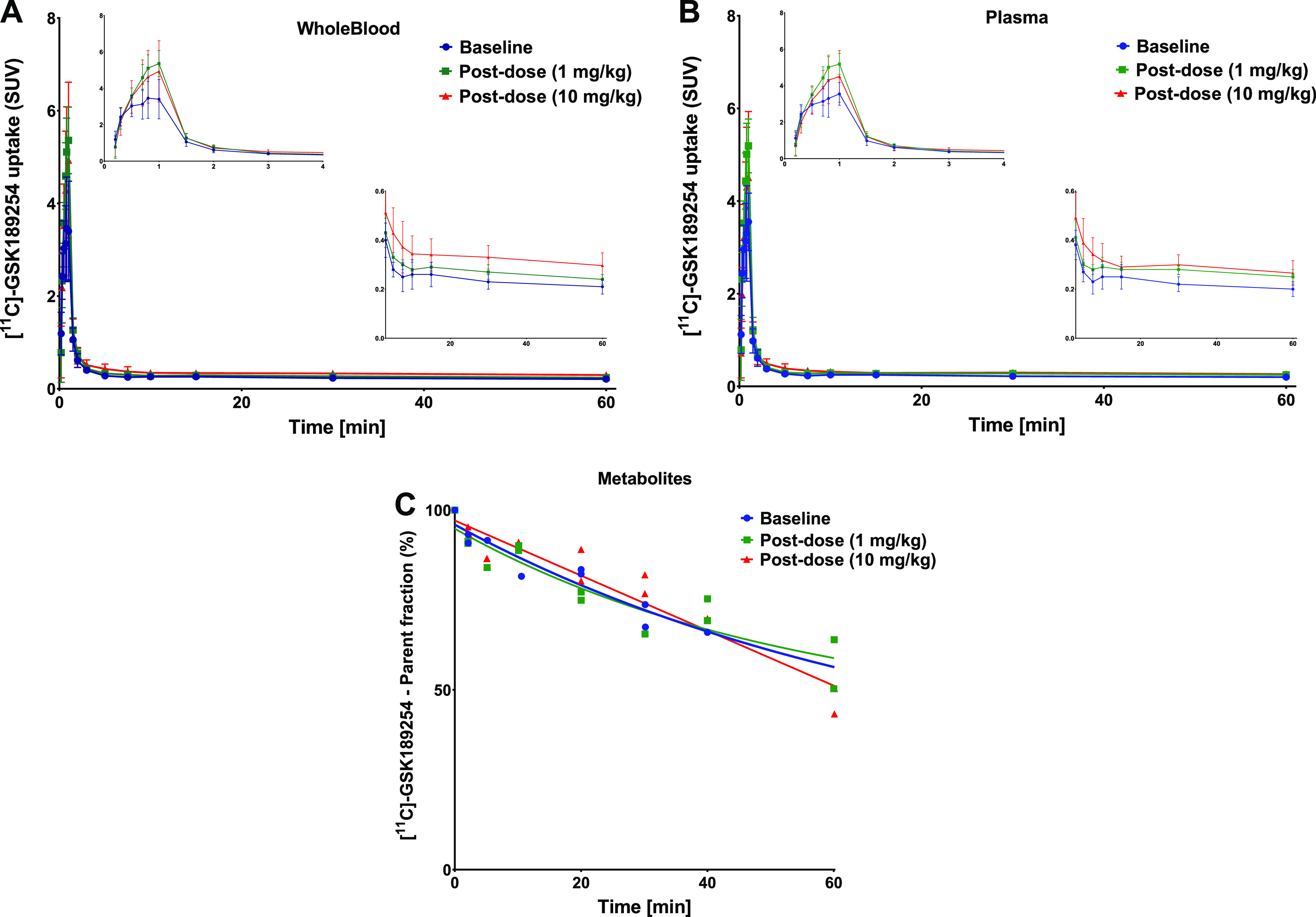
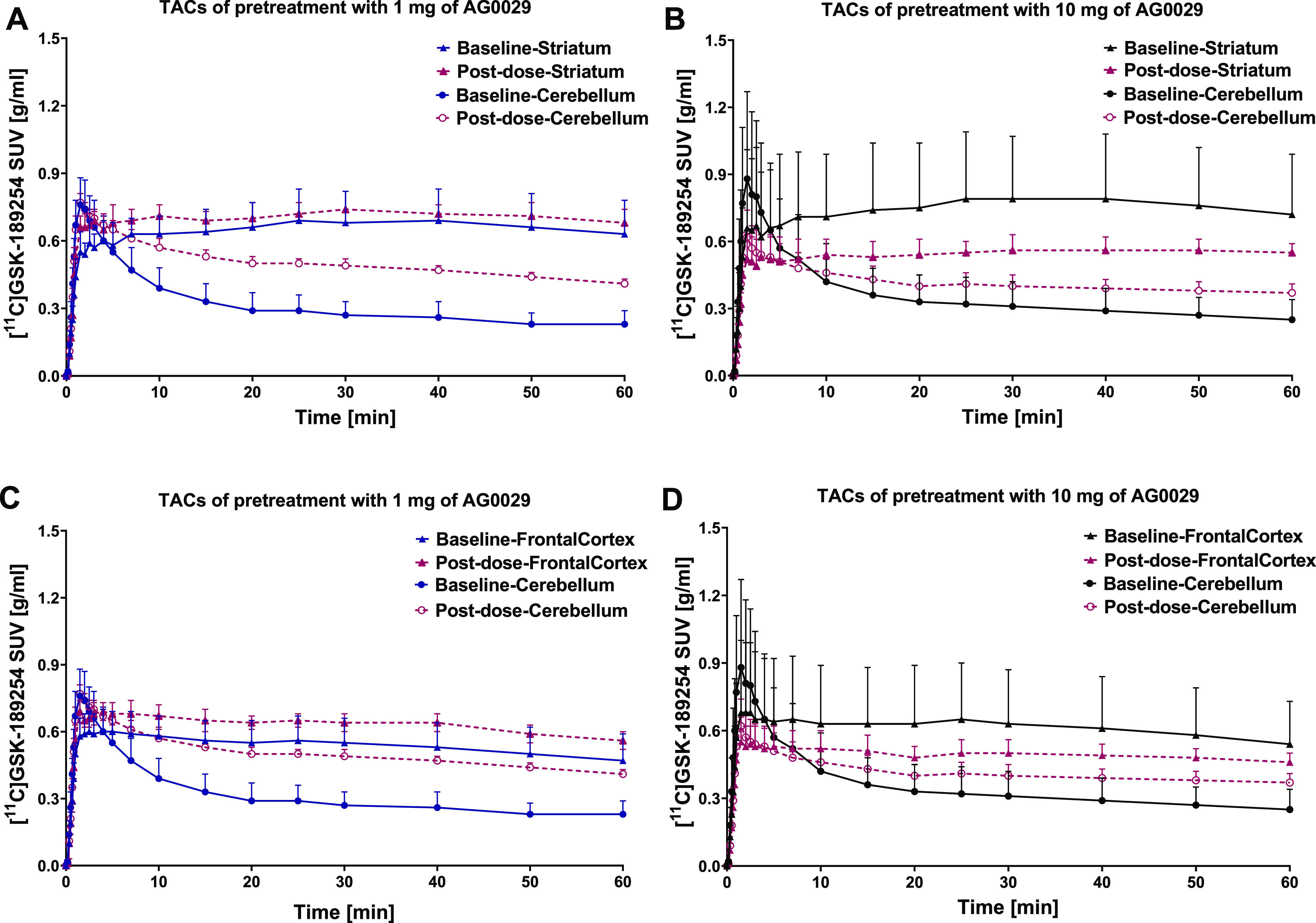
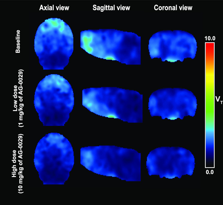
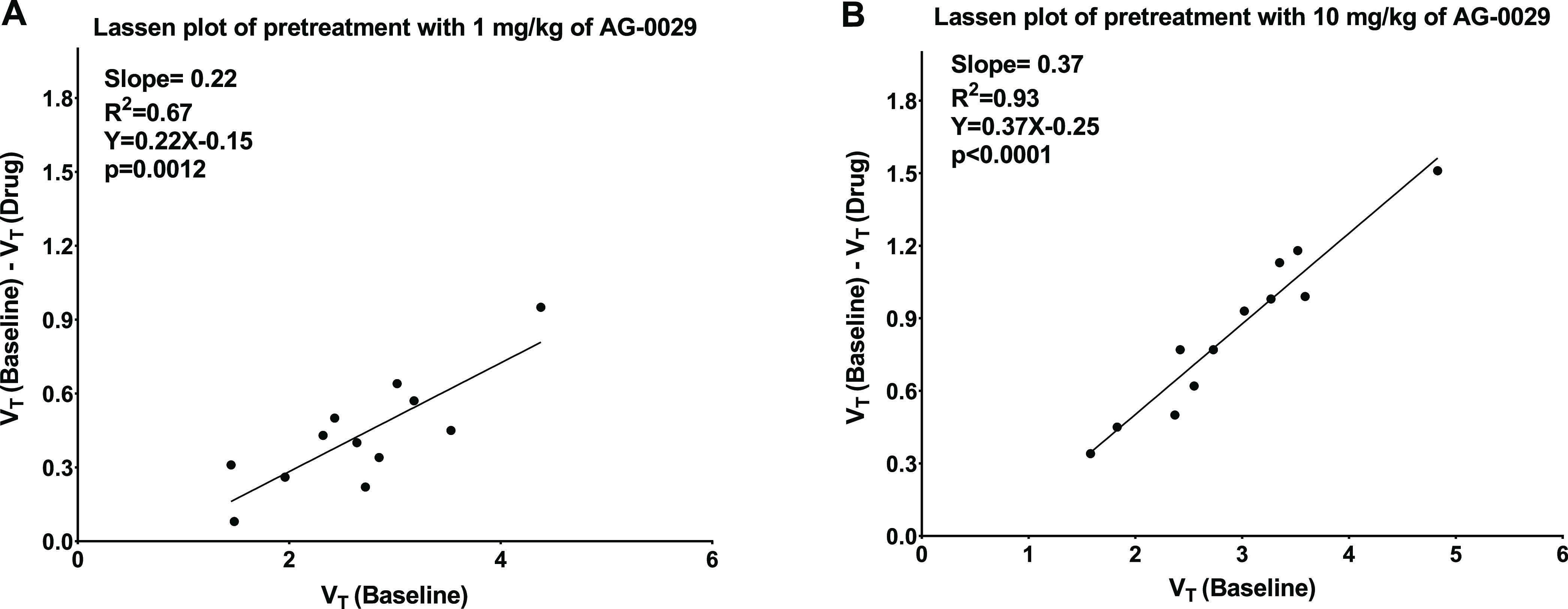
Introduction: Parkinson's disease (PD) is a neurodegenerative disorder characterized by motor dysfunction and a diverse range of nonmotor symptoms. Functional relationships between the dopaminergic and histaminergic systems suggest that dual-action pharmaceuticals like AG-0029 (D2/D3 agonist/H3 antagonist) could ameliorate both the motor and cognitive symptoms of PD. The current study aimed to demonstrate the interaction of AG-0029 with its intended targets in the mammalian brain using positron emission tomography (PET). Methods: Healthy male Wistar rats were scanned with a small-animal PET camera, using either the dopamine D2/D3 receptor ligand [11C]raclopride or the histamine H3 receptor ligand [11C]GSK-189254, before and after treatment with an intravenous, acute, single dose of AG-0029. Dynamic [11C]raclopride PET data (60 min duration) were analyzed using the simplified reference tissue model 2 (SRTM2) with cerebellum as reference tissue and the nondisplaceable binding potential as the outcome parameter. Data from dynamic [11C]GSK-189254 scans (60 min duration) with arterial blood sampling were analyzed using Logan graphical analysis with the volume of distribution (VT) as the outcome parameter. Receptor occupancy was estimated using a Lassen plot. Results: Dopamine D2/3 receptor occupancies in the striatum were 22.6 ± 18.0 and 84.0 ± 3.5% (mean ± SD) after administration of 0.1 and 1 mg/kg AG-0029, respectively. In several brain regions, the VT values of [11C]GSK-189254 were significantly reduced after pretreatment of rats with 1 or 10 mg/kg AG-0029. The H3 receptor occupancies were 11.9 ± 8.5 and 40.3 ± 11.3% for the 1 and 10 mg/kg doses of AG-0029, respectively. Conclusions: Target engagement of AG-0029 as an agonist at dopamine D2/D3 receptors and an antagonist at histamine H3 receptors could be demonstrated in the rat brain with [11C]raclopride and [11C]GSK-189254 PET, respectively. The measured occupancy values reflect the previously reported high (subnanomolar) affinity of AG-0029 to D2/D3 and moderate (submicromolar) affinity to H3 receptors.

Keywords: Parkinson’s disease; [11C]GSK-189254; [11C]raclopride; anti-Parkinson drug; dopamine D2 receptor; dual-action pharmaceutical; histamine H3 receptor; pharmacokinetic modeling.


Conflict of interest statement

The authors declare no competing financial interest.

Figures

Figure 1
Figure 1
Chemical structure of AG-0029.
Figure 2
Figure 2
Dose-dependent effect of AG-0029 administration on heart rate.
Figure 3
Figure 3
Average [11C]raclopride time–activity curves in the striatum (high D2 receptor expression) and cerebellum (low D2 receptor expression) at baseline and post-dose. Rats received either (A) a low dose (0.1 mg/kg) or (B) a high dose (1 mg/kg) of AG-0029. Data are plotted as mean ± SD.
Figure 4
Figure 4
Effect of AG-0029 on the influx (R1) of [11C]raclopride derived from SRTM2.
Figure 5
Figure 5
(A) Graphical comparison between baseline and post-dose [11C]raclopride binding potential (BPND) in rats pretreated with either 0.1 or 1 mg/kg of AG-0029 as the test drug. (B) Graphical presentation of the striatal D2 receptor occupancy versus the AG-0029 dose.
Figure 6
Figure 6
Two sets of BPND images of [11C]raclopride scans acquired at baseline (A, C) and after pretreatment with different doses of AG-0029 (B, D).
Figure 7
Figure 7
Kinetics of [11C]GSK-189254. Time–activity curves in (A) blood and (B) plasma at baseline and after administration of 1 or 10 mg/kg of AG-0029. The insets show the shape of the initial peak. (C) Parent fraction in plasma at baseline and post-dose. Error bars indicate standard deviation.
Figure 8
Figure 8
Representative time–activity curves (TACs) of [11C]GSK-182954 in the striatum and the cerebellum (A, B) and frontal cortex and the cerebellum (C, D) after pretreatment with a high (10 mg/kg) dose (B, D) and low (1 mg/kg) dose of AG-0029 (A, C).
Figure 9
Figure 9
Parametric (VT) images of [11C]GSK-189254 binding (axial, coronal, and sagittal views) of a representative rat brain at baseline (top row) and after administration of 1 mg/kg (middle row), or 10 mg/kg of AG-0029 (bottom row).
Figure 10
Figure 10
Examples of Lassen plots from (A) a rat that received a low dose (1 mg/kg) of AG-0029 and (B) a rat that received a high dose (10 mg/kg) of the drug before the post-dose [11C]GSK-189254 PET scan.

Similar articles

References

    1. Nussbaum R. L.; Ellis C. E. Alzheimer’s disease and Parkinson’s disease. N. Engl. J. Med. 2003, 348, 1356–1364. 10.1056/NEJM2003ra020003. - DOI - PubMed
    1. Schulz-Schaeffer W. J. The synaptic pathology of α-synuclein aggregation in dementia with Lewy bodies, Parkinson’s disease and Parkinson’s disease dementia. Acta Neuropathol. 2010, 120, 131–143. 10.1007/s00401-010-0711-0. - DOI - PMC - PubMed
    1. Villar-Piqué A.; Lopes da Fonseca T.; Outeiro T. F. Structure, function and toxicity of alpha-synuclein: the Bermuda triangle in synucleinopathies. J. Neurochem. 2016, 139, 240–255. 10.1111/jnc.13249. - DOI - PubMed
    1. Burré J.; Sharma M.; Südhof T. C. Cell biology and pathophysiology of α-synuclein. Cold Spring Harbor Perspect. Med. 2018, 8, a024091 10.1101/cshperspect.a024091. - DOI - PMC - PubMed
    1. Kalia L. V.; Lang A. E. Parkinson’s disease. Lancet 2015, 386, 896–912. 10.1016/S0140-6736(14)61393-3. - DOI - PubMed