. 2022 Aug 4;82(15):2858-2870.e8.
doi: 10.1016/j.molcel.2022.05.025. Epub 2022 Jun 21.

Supramolecular assembly of GSK3α as a cellular response to amino acid starvation

Affiliations

Supramolecular assembly of GSK3α as a cellular response to amino acid starvation

Laura Hinze et al. Mol Cell. .

Abstract

The tolerance of amino acid starvation is fundamental to robust cellular fitness. Asparagine depletion is lethal to some cancer cells, a vulnerability that can be exploited clinically. We report that resistance to asparagine starvation is uniquely dependent on an N-terminal low-complexity domain of GSK3α, which its paralog GSK3β lacks. In response to depletion of specific amino acids, including asparagine, leucine, and valine, this domain mediates supramolecular assembly of GSK3α with ubiquitin-proteasome system components in spatially sequestered cytoplasmic bodies. This effect is independent of mTORC1 or GCN2. In normal cells, GSK3α promotes survival during essential amino acid starvation. In human leukemia, GSK3α body formation predicts asparaginase resistance, and sensitivity to asparaginase combined with a GSK3α inhibitor. We propose that GSK3α body formation provides a cellular mechanism to maximize the catalytic efficiency of proteasomal protein degradation in response to amino acid starvation, an adaptive response co-opted by cancer cells for asparaginase resistance.

Keywords: GSK3; Wnt; asparaginase; protein degradation; ubiquitin-proteasome system.


Conflict of interest statement

Declaration of interests Boston Children’s Hospital has filed patents on the subject matter of this manuscript. G.C. is a founder of Samus Therapeutics and a member of its board of directors. A.G. is on a scientific advisory board for Attivare Therapeutics and receives research funding from Astellas Pharma.

Figures
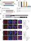
Figure 1.
Figure 1.. Resistance to asparaginase requires an N-terminal low-complexity domain of GSK3α that triggers its spatial sequestration.
(A) Protein sequences of human GSK3α (uniprot ID P49840) and GSK3β (uniprot P49841) were aligned and conservation was assessed using ClustalW. (B) Jurkat cells were transduced with the indicated constructs, treated with asparaginase for 8 days, and relative viability was assessed. Unless indicated by brackets, all significance assessments reflect comparisons to the shLuc control. Statistical significance assessed by a one-way ANOVA with Dunnett’s adjustment for multiple comparisons. (C) Prediction of low-complexity SEG domains was performed for the indicated proteins [as in (A)] using DisMeta analysis. The kinase and AXIN-binding domains are shown for reference. Domain boundaries from the uniprot identifiers in (A). (D) ClustalW alignment of the N-terminal domains of human GSK3α and GSK3β. (E) Jurkat cells were treated with vehicle or 100 U/L asparaginase for 48 hours, and GSK3α localization was assessed by confocal immunofluorescence microscopy. DAPI served as the nuclear stain. Images are representatives from at least 3 biologic replicates. For GSK3α body quantification, GSK3α positive puncta were counted in 50 cells in biologic triplicates. Statistical significance was assessed by a two-sided Student’s t-test with Welch adjustment. (F) Jurkat cells were treated as described in (E), and GSK3β localization was assessed using confocal immunofluorescence microscopy. GSK3β body counting and statistical significance was assessed as in (E). All error bars represent SEM. Scale bars, 5 µm or 2 µm (inset). **** p ≤ 0.0001, *** p ≤ 0.001., ** p ≤ 0.01, * p < 0.05, n.s. p ≥ 0.05. See also Figure S1–S3.
Figure 2.
Figure 2.. Asparagine starvation triggers spatial sequestration of GSK3α into membraneless cytoplasmic bodies.
(A) Jurkat cells were treated with vehicle, asparaginase (100 U/L), dexamethasone (1 µM), vincristine (10 nM), or mercaptopurine (1 µM) for 48 hours. GSK3α body formation was assessed by confocal immunofluorescence analysis for GSK3α. GSK3α body quantification was performed by counting the number of GSK3α positive puncta in 50 cells in biologic triplicates. Statistical significance was assessed using a one-way ANOVA with Dunnett’s adjustment. (B) Jurkat cells were cultured in RPMI medium containing only essential amino acids, with the addition of either vehicle, asparagine (0.37 mM), or glutamine (2 mM) in biological triplicates. After 48 hours, quantification of GSK3α bodies was assessed as in (A). Statistical significance was assessed by a one-way ANOVA with Dunnett’s adjustment. (C) Representative images of the experiment described in (B). Scale bars, 5 µm or 2 µm (inset). (D) Jurkat cells were transduced with expression constructs encoding the N-terminus of either GSK3α (amino acids 1–118) or GSK3β (amino acids 1–55) fused to HaloTag. Cells were treated with 100 U/L asparaginase for 48 hrs, and stained with the fluorescent ligand Oregon Green to detect HaloTag positive bodies. Confocal microscopy was the performed, and HaloTag body quantification were performed as described in (A). Statistical significance was assessed by a two-sided Student’s t-test with Welch adjustment. All error bars represent SEM. **** p ≤ 0.0001, *** p ≤ 0.001., ** p ≤ 0.01, * p < 0.05, n.s. p ≥ 0.05.
Figure 3.
Figure 3.. Asparagine depletion triggers supramolecular assembly of GSK3α with ubiquitin-proteasome system components.
(A) Jurkat cells were treated with vehicle, asparaginase (100 U/L), BRD0705 (1 µM) or both drugs in combination (combo) for 48 hours. Cells were fixed and stained with primary antibodies against GSK3α and ubiquitin, and secondary Alexa-Fluor 488 and Alexa-Fluor 555 antibodies. DAPI served as the nuclear stain. Images are representatives from at least 3 biologic replicates. (B) Puncta positive for GSK3α, ubiquitin, or both markers colocalized were counted in 50 cells in biological triplicates in cells from the experiment shown in (A). Significance was assessed by one-way ANOVA with Dunnett’s adjustment for multiple comparisons. (C) Jurkat cells were treated with vehicle or a 100 U/L asparaginase dose for 48 hours, and confocal immunofluorescence microscopy for GSK3α and the proteasomal subunit PSMA4 was performed. (D) Puncta positive for GSK3α, PSMA4, or both markers colocalized were counted and significance was assessed as in (B). (E) Jurkat cells were treated with vehicle or sucrose (0.2 M) for 48 hours. GSK3α localization was assessed by confocal immunofluorescence microscopy. The number of foci positive for GSK3α was counted in 50 cells in biological triplicates from each condition. Significance was assessed by a two-sided Student’s t-test with Welch adjustment. All error bars represent SEM. Scale bars, 5 µm or 2 µm (inset). **** p ≤ 0.0001, *** p ≤ 0.001., ** p ≤ 0.01, * p < 0.05, n.s. p ≥ 0.05. See also Figure S4–S6 and Table S2.
Figure 4.
Figure 4.. HSP70 is required for GSK3α body formation and asparaginase resistance.
(A) Jurkat cells were treated with vehicle or 100 U/L asparaginase for 48 hrs, and analyzed by confocal immunofluorescence microscopy for GSK3α and HSP70. Images are representative of at least 3 biological replicates. (B) Bodies individually positive for GSK3α, HSP70, or both markers colocalized in cells from (A) were counted in 50 cells in biologic triplicates. Significance was assessed using a one-way ANOVA with Dunnett’s adjustment. (C) Jurkat cells were treated with vehicle or asparaginase for 48hrs in the presence of the HSP70 inhibitors 27c or B (5 µM). GSK3α body formation was assessed by confocal immunofluorescence microscopy as in (A-B). (D) Jurkat cells were transduced with shLuciferase or shHSP70, and treated with vehicle or 100 U/L asparaginase for 48 hours. GSK3α body formation was assessed as in (C). Significance was determined by one-way ANOVA with Dunnett’s adjustment. (E) Jurkat cells were treated with the indicated drugs for 4 days in biological triplicates, and viability was assessed by trypan blue viable cell counts. All cell counts are normalized to those in vehicle-treated cells. (F) Jurkat cells were transduced with shLuciferase or with the indicated HSP70 shRNAs, treated with the indicated doses of asparaginase for 4 days, and viability was assessed as in (E). All cell counts are normalized to those in shLuc vehicle treated cells. (G) Jurkat cells were treated as in (A), and then stained for antibodies for GSK3α and the second heat shock protein indicated. Formation of bodies positive for each individual marker and for both markers colocalized was assessed as in (B). (H) Jurkat cells were treated with vehicle or the HSP90 inhibitor geldanamycin (500 nM), together with vehicle or asparaginase (100 U/L). GSK3α body formation was assessed as in (B). (I) Jurkat cells were treated as in (H), and viability was assessed as in (E). All error bars represent SEM. Scale bars, 5 µm or 2 µm (inset). **** p ≤ 0.0001, *** p ≤ 0.001., ** p ≤ 0.01, * p < 0.05, n.s. p ≥ 0.05. See also Figure S7–S9.
Figure 5.
Figure 5.. GSK3α promotes survival during essential amino acid starvation.
(A) Jurkat cells were cultured in complete RPMI medium or in RPMI lacking all essential amino acids for 48 hrs, and GSK3α body formation was assessed by confocal immunofluorescence microscopy in biological triplicates. Statistical significance was assessed by a two-sided Student’s t-test with Welch adjustment. (B) Jurkat cells were cultured in complete RPMI, or in medium individually deficient in the indicated amino acids. GSK3α body formation was assessed in biological duplicates. Significance was assessed by a one-way ANOVA with Dunnett adjustment for multiple comparisons. (C) Jurkat cells were cultured in complete RPMI, or in medium individually deficient in arginine. GSK3α body formation was assessed as in (B). Significance was assessed by a two-sided Student’s t-test with Welch adjustment. (D) Jurkat cells were cultured in leucine- or valine-deficient medium in biological triplicates, together with vehicle or the GSK3α-selective inhibitor BRD0705 (1 µM) for 48 hrs, without or with supplementation of the indicated amino acids. Viability is normalized to vehicle-treated cells. Significance was assessed by a one-way ANOVA with Dunnett adjustment for multiple comparisons. (E) Normal human CD34+ hematopoietic progenitors were cultured in complete RPMI, or in medium lacking all essential amino acids for 48 hours. GSK3α body formation was assessed in biological duplicates. Significance was assessed by two-sided Student’s t-test with Welch adjustment. (F) Normal CD34+ human hematopoietic progenitors were cultured in RPMI, or in RPMI deficient in essential amino acids in the presence of vehicle, the GSK3α-selective inhibitor BRD0705, or its enantiomeric negative control BRD5648. At 48 hours, viability was assessed in biological duplicates. Significance was assessed as in (D). (G) CCD841 cells were cultured in complete DMEM or in DMEM lacking all essential amino acids for 48 hours. GSK3α body formation and significance were assessed as in (E). (H) CCD841 cells were cultured in complete or amino acid deficient medium as in (G), treated with the indicated drugs as in (F), and relative viability and statistical significance were assessed as in (F). All error bars represent SEM. **** p ≤ 0.0001, *** p ≤ 0.001., ** p ≤ 0.01, * p < 0.05, n.s. p ≥ 0.05. See als Figure S9.
Figure 6.
Figure 6.. GSK3α body formation predicts asparaginase response in human leukemia.
(A) Leukemia cell lines (Table S3) were treated with vehicle or asparaginase (100 U/L) in biological duplicates for 48 hours, and GSK3α body formation was assessed by confocal immunofluorescence microscopy. Cells were classified as asparaginase-sensitive vs. resistant as shown in Figure S10A. Statistical significance was assessed by a two-sided Student’s t-test with Welch adjustment. (B) Patient-derived xenograft (PDX) leukemia specimens (Table S4) were treated with asparaginase in biological duplicates and GSK3α body formation was assessed as in (A). Cells were classified as asparaginase-sensitive or resistant as shown in Figure S10B. Statistical significance was assessed as in (A). (C) Receiver operating characteristic (ROC) analysis of GSK3α body formation as a biomarker of asparaginase response from the experiments shown in (A-B). (D) Assessment of GSK3α body formation after treatment with asparaginase for 48 hrs as in (A), in PDX models collected at initial diagnosis from patients who had an excellent response to asparaginase-intensive combination chemotherapy, versus those collected at relapse. Statistical significance was assessed by a two-sided Student’s t-test with Welch adjustment. (E) Human leukemia cell lines (Table S3) were treated with vehicle or combo (asparaginase 100 U/L and BRD0705 1 µM) in biological duplicates, and GSK3α body formation was assessed as in (A). Cell lines were classified as combo sensitive or resistant based on the viability data shown in Figure S10C. Significance was assessed as in (A). (F) Human leukemia PDX samples (Table S4) were treated in biological duplicates and GSK3α body formation was assessed as in (E). PDX samples were classified as combo-sensitive versus resistant based on the viability data shown in Figure S10D. Significance was assessed as in (A). (G) ROC analysis human leukemia cell lines and PDXs from the experiments shown in (E-F). In A, B, E, F each data point represents the average of biologic duplicates from an individual cell line or PDX model. All error bars represent SEM. **** p ≤ 0.0001, *** p ≤ 0.001., ** p ≤ 0.01, * p < 0.05, n.s. p ≥ 0.05. See also Figure S10 and Table S3–S4.
Figure 7.
Figure 7.. Proposed Model.
Depletion of any of the indicated amino acids is sufficient to stimulate supramolecular assembly of GSK3α with components of the ubiquitin proteasome system in spatially sequestered cytoplasmic bodies. We propose that this provides a cellular mechanism to maximize the catalytic activity of proteasomal protein degradation in response to amino acid starvation by concentrating its enzymatic components within GSK3α bodies.

References

    1. Acebron SP, Karaulanov E, Berger BS, Huang YL, and Niehrs C (2014). Mitotic wnt signaling promotes protein stabilization and regulates cell size. Mol Cell 54, 663–674. - PubMed
    1. Ahmad F, and Woodgett JR (2020). Emerging roles of GSK-3alpha in pathophysiology: Emphasis on cardio-metabolic disorders. Biochim Biophys Acta Mol Cell Res 1867, 118616. - PubMed
    1. An H, Ordureau A, Korner M, Paulo JA, and Harper JW (2020). Systematic quantitative analysis of ribosome inventory during nutrient stress. Nature 583, 303–309. - PMC - PubMed
    1. Appel IM, den Boer ML, Meijerink JP, Veerman AJ, Reniers NC, and Pieters R (2006). Up-regulation of asparagine synthetase expression is not linked to the clinical response L-asparaginase in pediatric acute lymphoblastic leukemia. Blood 107, 4244–4249. - PubMed
    1. Banani SF, Lee HO, Hyman AA, and Rosen MK (2017). Biomolecular condensates: organizers of cellular biochemistry. Nature reviews Molecular cell biology 18, 285–298. - PMC - PubMed

Publication types