Review
. 2022 Jul;36(7):1720-1748.
doi: 10.1038/s41375-022-01620-2. Epub 2022 Jun 22.

The 5th edition of the World Health Organization Classification of Haematolymphoid Tumours: Lymphoid Neoplasms

Rita Alaggio  1 Catalina Amador  2 Ioannis Anagnostopoulos  3 Ayoma D Attygalle  4 Iguaracyra Barreto de Oliveira Araujo  5 Emilio Berti  6 Govind Bhagat  7 Anita Maria Borges  8 Daniel Boyer  9 Mariarita Calaminici  10 Amy Chadburn  11 John K C Chan  12 Wah Cheuk  12 Wee-Joo Chng  13 John K Choi  14 Shih-Sung Chuang  15 Sarah E Coupland  16 Magdalena Czader  17 Sandeep S Dave  18 Daphne de Jong  19 Ming-Qing Du  20 Kojo S Elenitoba-Johnson  21 Judith Ferry  22 Julia Geyer  11 Dita Gratzinger  23 Joan Guitart  24 Sumeet Gujral  25 Marian Harris  26 Christine J Harrison  27 Sylvia Hartmann  28 Andreas Hochhaus  29 Patty M Jansen  30 Kennosuke Karube  31 Werner Kempf  32 Joseph Khoury  33 Hiroshi Kimura  34 Wolfram Klapper  35 Alexandra E Kovach  36 Shaji Kumar  37 Alexander J Lazar  38 Stefano Lazzi  39 Lorenzo Leoncini  39 Nelson Leung  40 Vasiliki Leventaki  41 Xiao-Qiu Li  42 Megan S Lim  21 Wei-Ping Liu  43 Abner Louissaint Jr  44 Andrea Marcogliese  45 L Jeffrey Medeiros  33 Michael Michal  46 Roberto N Miranda  33 Christina Mitteldorf  47 Santiago Montes-Moreno  48 William Morice  49 Valentina Nardi  44 Kikkeri N Naresh  50 Yasodha Natkunam  23 Siok-Bian Ng  51 Ilske Oschlies  35 German Ott  52 Marie Parrens  53 Melissa Pulitzer  54 S Vincent Rajkumar  55 Andrew C Rawstron  56 Karen Rech  49 Andreas Rosenwald  3 Jonathan Said  57 Clémentine Sarkozy  58 Shahin Sayed  59 Caner Saygin  60 Anna Schuh  61 William Sewell  62 Reiner Siebert  63 Aliyah R Sohani  44 Reuben Tooze  64 Alexandra Traverse-Glehen  65 Francisco Vega  33 Beatrice Vergier  66 Ashutosh D Wechalekar  67 Brent Wood  36 Luc Xerri  68 Wenbin Xiao  54
Affiliations
Review

The 5th edition of the World Health Organization Classification of Haematolymphoid Tumours: Lymphoid Neoplasms

Rita Alaggio et al. Leukemia. 2022 Jul.

Erratum in

  • Correction: "The 5th edition of The World Health Organization Classification of Haematolymphoid Tumours: Lymphoid Neoplasms" Leukemia. 2022 Jul;36(7):1720-1748.
    Alaggio R, Amador C, Anagnostopoulos I, Attygalle AD, de Oliveira Araujo IB, Berti E, Bhagat G, Borges AM, Boyer D, Calaminici M, Chadburn A, Chan JKC, Cheuk W, Chng WJ, Choi JK, Chuang SS, Coupland SE, Czader M, Dave SS, de Jong D, Di Napoli A, Du MQ, Elenitoba-Johnson KS, Ferry J, Geyer J, Gratzinger D, Guitart J, Gujral S, Harris M, Harrison CJ, Hartmann S, Hochhaus A, Jansen PM, Karube K, Kempf W, Khoury J, Kimura H, Klapper W, Kovach AE, Kumar S, Lazar AJ, Lazzi S, Leoncini L, Leung N, Leventaki V, Li XQ, Lim MS, Liu WP, Louissaint A Jr, Marcogliese A, Medeiros LJ, Michal M, Miranda RN, Mitteldorf C, Montes-Moreno S, Morice W, Nardi V, Naresh KN, Natkunam Y, Ng SB, Oschlies I, Ott G, Parrens M, Pulitzer M, Rajkumar SV, Rawstron AC, Rech K, Rosenwald A, Said J, Sarkozy C, Sayed S, Saygin C, Schuh A, Sewell W, Siebert R, Sohani AR, Suzuki R, Tooze R, Traverse-Glehen A, Vega F, Vergier B, Wechalekar AD, Wood B, Xerri L, Xiao W; International Agency for Research on Cancer/World Health Organization. Alaggio R, et al. Leukemia. 2023 Sep;37(9):1944-1951. doi: 10.1038/s41375-023-01962-5. Leukemia. 2023. PMID: 37468552 Free PMC article. No abstract available.

Abstract

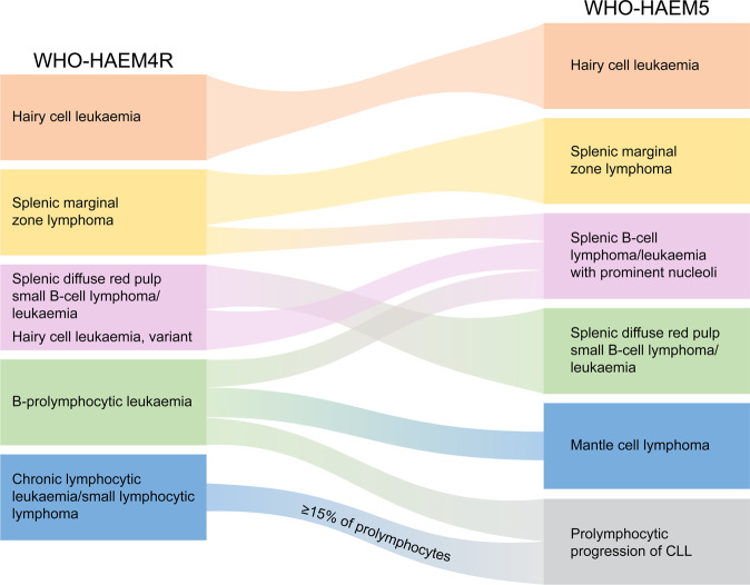
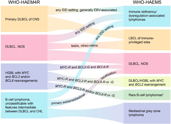
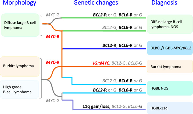
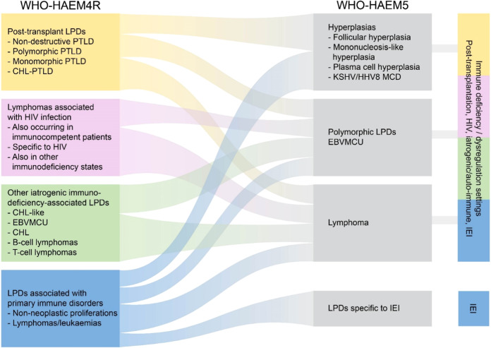
We herein present an overview of the upcoming 5th edition of the World Health Organization Classification of Haematolymphoid Tumours focussing on lymphoid neoplasms. Myeloid and histiocytic neoplasms will be presented in a separate accompanying article. Besides listing the entities of the classification, we highlight and explain changes from the revised 4th edition. These include reorganization of entities by a hierarchical system as is adopted throughout the 5th edition of the WHO classification of tumours of all organ systems, modification of nomenclature for some entities, revision of diagnostic criteria or subtypes, deletion of certain entities, and introduction of new entities, as well as inclusion of tumour-like lesions, mesenchymal lesions specific to lymph node and spleen, and germline predisposition syndromes associated with the lymphoid neoplasms.


Conflict of interest statement

The authors declare no competing interests.

Figures

Fig. 1
Fig. 1. Summary of the relationship between splenic B-cell lymphoma entities as named and defined in the revised 4th edition of the WHO classification (WHO-HAEM4R) and in the present 5th edition (WHO-HAEM5).
Some cases previously classified as B-prolymphocytic leukaemia do represent (blastoid) mantle cell lymphoma (as was already indicated in WHO-HAEM4R) or prolymphocytic progression of CLL. Cases classified in WHO-HAEM4R as CLL/SLL with ≥ 15% of prolymphocytes are now classified as prolymphocytic progression of CLL, cases with <15% of prolymphocytes remain CLL/SLL in WHO-HAEM5. Remaining cases are now renamed as “splenic B-cell lymphoma/leukaemia with prominent nucleoli” (SBLPN). This latter entity has absorbed cases formerly classified as hairy cell leukaemia variant (HCLv) and very rare cases of splenic marginal zone lymphoma with similar morphological features. It should be noted that the distinction between the various entities cannot always be made in the absence of a splenectomy specimen.
Fig. 2
Fig. 2. Aetiology and recurrent genetic abnormalities in extranodal marginal zone lymphoma (EMZL) of various sites.
An important clinical application is that BIRC3::MALT1 identifies those cases of the gastric EMZL not responding to H. pylori eradication. As many of the genes involved in EMZL have not been uniformly investigated across different sites, only the recurrent genetic changes fundamental to the understanding of EMZL pathogenesis are presented. The height of the boxes under sites does not reflect the frequencies of these lymphomas. trans translocation, mut mutation, del: deletion.
Fig. 3
Fig. 3. Summary of the relationship between large B-cell lymphoma (LBCL) entities as named and defined in the revised 4th edition of the WHO classification (WHO-HAEM4R) and in the present 5th edition (WHO-HAEM5).
* “Rare B-cell lymphomas” refer to those fulfilling definitions of specific clinico-pathological entities while incidentally bearing concomitant MYC and BCL2 rearrangements. Examples are fluid-overload-associated large B-cell lymphomas and rare follicular lymphomas. R rearrangement, G germline configuration.
Fig. 4
Fig. 4. Algorithm for classification of aggressive B-cell lymphomas in WHO-HAEM5 in the light of MYC, BCL2 and BCL6 rearrangement and complex 11q gain/loss patterns.
HGBL high grade B-cell lymphoma, R rearrangement, G germline configuration.
Fig. 5
Fig. 5. Summary of the relationship between immunodeficiency-associated lymphoid proliferations and lymphomas as named and defined in the revised 4th edition of the WHO Classification (WHO-HAEM4R) and in the present 5th edition (WHO-HAEM5).
The overarching concept applied in WHO-HAEM5 recognizes the pathological and biological similarities between proliferations presenting in various immune deficiency settings, while acknowledging their specific features. Outside the shared entities, unique proliferations are especially typical for various inborn errors of immunity (IEI). EBVMCU: EBV-positive mucocutaneous ulcer.
Fig. 6
Fig. 6. Indolent NK-cell lymphoproliferative disorder of the gastrointestinal tract involving the stomach.
The gastric mucosa shows expansion of the lamina propria by an atypical lymphoid infiltrate. The tumour cells are medium-sized, often with pale-staining cytoplasm.
Fig. 7
Fig. 7. Nodal TFH-cell lymphoma (nTFHL).
A Nodal TFH-cell lymphoma, angioimmunoblastic-type (nTFHL-AI). The normal architecture of the lymph node is effaced. There is a diffuse infiltrate of medium-sized, slightly atypical lymphocytes, sometimes with clear cytoplasm. One of the hallmarks of the disease is the proliferation of arborizing post-capillary vessels consistent with high endothelial venules. B Nodal TFH-cell lymphoma, follicular-type (nTFHL-F). In this example, progressive transformation of germinal centre-like nTFHL-F, clusters of atypical lymphoid cells with pale cytoplasm are embedded in a background of small lymphocytes of mantle zone type. The inset shows strong expression of PD1 in the tumour cells. C Nodal TFH-cell lymphoma, not otherwise specified. This tumour is composed of a sheet-like proliferation of medium-sized to large neoplastic cells.
Fig. 8
Fig. 8. Nodal EBV-positive T- and NK-cell lymphoma.
This lymphoma shows a diffuse infiltrate of relatively monotonous, medium-sized to large cells, sometimes reminiscent of centroblasts. Inset: in-situ hybridization for EBERs identifies EBV infection in virtually all tumour cells.

Comment in

Similar articles

Cited by

References

    1. Jaffe ES, Harris N, Stein H, Vardiman JW (Eds.): World Health Organization classification of Tumours. Pathology and Genetics of Tumours of Haematopoietic and Lymphoid Tissues. 3rd ed. Lyon: IARC; 2001.
    1. Harris NL, Jaffe ES, Stein H, Banks PM, Chan JK, Cleary ML, et al. A revised European-American classification of lymphoid neoplasms: a proposal from the International Lymphoma Study Group. Blood. 1994;84:1361–92. - PubMed
    1. Swerdlow SH, Campo E, Harris NL, Jaffe ES, Pileri SA, Stein H, Thiele J, et al. (Eds.): World Health Organization classification of Tumours of Haematopoietic and Lymphoid Tissues. 4th ed. Lyon: IARC 2008.
    1. Swerdlow SH, Campo E, Harris NL, Jaffe ES, Pileri SA, Stein H, Thiele J (Eds.): World Health Organization classification of Tumours of Haematopoietic and Lymphoid Tissues. Revised 4th ed. Lyon: IARC; 2017.
    1. Fajgenbaum DC, Uldrick TS, Bagg A, Frank D, Wu D, Srkalovic G, et al. International, evidence-based consensus diagnostic criteria for HHV-8-negative/idiopathic multicentric Castleman disease. Blood. 2017;129:1646–57. - PMC - PubMed