

Potential biomarkers of spinal dural arteriovenous fistula: C4BPA and C1QA
- PMID: 35733178
- PMCID: PMC9215050
- DOI: 10.1186/s12974-022-02522-x
Potential biomarkers of spinal dural arteriovenous fistula: C4BPA and C1QA
Abstract
Background and purpose: A major challenge in spinal dural arteriovenous fistula (SDAVF) is timely diagnosis, but no specific predictive biomarkers are known.
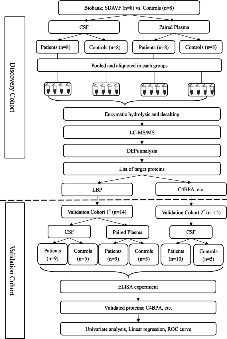
Methods: In the discovery cohort (case, n = 8 vs. control, n = 8), we used cerebrospinal fluid (CSF) and paired plasma samples to identify differentially expressed proteins by label-free quantitative proteomics. Further bioinformatics enrichment analyses were performed to screen target proteins. Finally, it was validated by ELISA in two of the new cohorts (case, n = 17 vs. control, n = 9), and univariate analysis, simple linear regression, and receiver operator characteristic (ROC) curve analysis were performed to evaluate the diagnostic potential.
Results: In the discovery cohort, the most overexpressed proteins were APOB and C4BPA in CSF samples of patients. The GO/KEGG enrichment analysis indicated that the upregulated proteins were mainly involved in the acute inflammatory response and complement activation. Hub-gene analysis revealed that APP might be the key protein in the molecular interaction network. In the validation cohort, C4BPA and C1QA were significantly overexpressed in the CSF of patients, averaging 3046.9 ng/ml and 2167.2 ng/ml, respectively. Simple linear regression demonstrated that levels of C1QA and C4 were positively correlated with total protein in CSF (R2 = 0.8021, p = 0.0005; R2 = 0.7447, p = 0.0013). The areas under the ROC curves of C4BPA and C1QA were 0.86 and 1.00, respectively.
Conclusions: This study was the first to identify C4BPA and C1QA as potential biomarkers for the diagnosis of SDAVF and revealed that complement pathway activation might be one of the molecular mechanisms for venous hypertension myelopathy.
Keywords: Biomarkers; C1QA; C4BPA; Spinal dural arteriovenous fistula; Venous hypertension myelopathy.
© 2022. The Author(s).
Conflict of interest statement
The authors declare that they have no competing interests.
Figures


Similar articles
-
Identification of a novel serum biomarker for pancreatic cancer, C4b-binding protein α-chain (C4BPA) by quantitative proteomic analysis using tandem mass tags.Br J Cancer. 2016 Oct 11;115(8):949-956. doi: 10.1038/bjc.2016.295. Epub 2016 Sep 22. Br J Cancer. 2016. PMID: 27657339 Free PMC article.
-
[Spinal dural arteriovenous fistula presented with rapidly progressive myelopathy, longitudinally extensive spinal cord lesion, pleocytosis with polymorphonuclear predominance, and decreased cerebrospinal fluid glucose levels: a case report].Rinsho Shinkeigaku. 2020 Oct 24;60(10):699-705. doi: 10.5692/clinicalneurol.cn-001472. Epub 2020 Sep 5. Rinsho Shinkeigaku. 2020. PMID: 32893247 Review. Japanese.
-
A simple score (AVFS) to identify spinal dural arteriovenous fistula before spinal digital subtraction angiography.J Stroke Cerebrovasc Dis. 2014 Sep;23(8):1995-2000. doi: 10.1016/j.jstrokecerebrovasdis.2014.01.021. Epub 2014 Aug 6. J Stroke Cerebrovasc Dis. 2014. PMID: 25106829
-
Quantitative proteomics revealed novel proteins associated with molecular subtypes of breast cancer.J Proteomics. 2016 Oct 4;148:183-93. doi: 10.1016/j.jprot.2016.07.033. Epub 2016 Aug 3. J Proteomics. 2016. PMID: 27498393
-
Spinal Dural Arteriovenous Fistula: A Review.Adv Tech Stand Neurosurg. 2016;(43):111-37. doi: 10.1007/978-3-319-21359-0_5. Adv Tech Stand Neurosurg. 2016. PMID: 26508408 Review.
Cited by
-
New insight into DAVF pathology-Clues from meningeal immunity.Front Immunol. 2022 Sep 15;13:858924. doi: 10.3389/fimmu.2022.858924. eCollection 2022. Front Immunol. 2022. PMID: 36189220 Free PMC article. Review.
-
Peripheral macrophages in the development and progression of structural cerebrovascular pathologies.J Cereb Blood Flow Metab. 2024 Feb;44(2):169-191. doi: 10.1177/0271678X231217001. Epub 2023 Nov 24. J Cereb Blood Flow Metab. 2024. PMID: 38000039 Free PMC article. Review.
-
The Complement System and C4b-Binding Protein: A Focus on the Promise of C4BPα as a Biomarker to Predict Clopidogrel Resistance.Mol Diagn Ther. 2024 Mar;28(2):189-199. doi: 10.1007/s40291-023-00691-w. Epub 2024 Jan 23. Mol Diagn Ther. 2024. PMID: 38261250 Review.
-
Rupture of a Spinal Dural Arteriovenous Fistula as a Differential Diagnosis of a Coronary Syndrome: Case Report.Neurosurg Pract. 2023 Aug 11;4(3):e00050. doi: 10.1227/neuprac.0000000000000050. eCollection 2023 Sep. Neurosurg Pract. 2023. PMID: 39958797 Free PMC article.
References
MeSH terms
Substances
Grants and funding
LinkOut - more resources
Full Text Sources
Medical
Molecular Biology Databases
Miscellaneous
