

Emerging Insight Into the Role of Circadian Clock Gene BMAL1 in Cellular Senescence
- PMID: 35733785
- PMCID: PMC9207346
- DOI: 10.3389/fendo.2022.915139
Emerging Insight Into the Role of Circadian Clock Gene BMAL1 in Cellular Senescence
Abstract
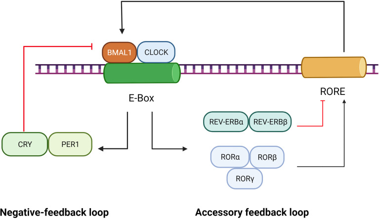
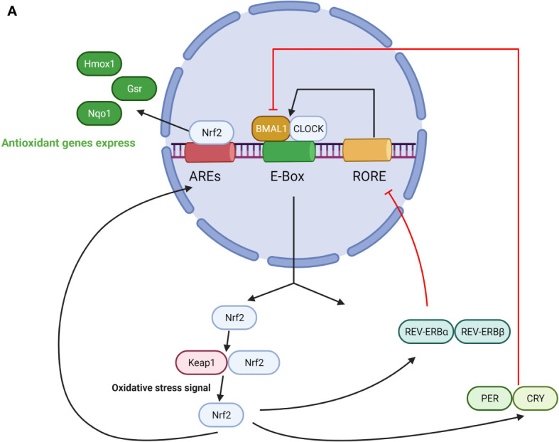
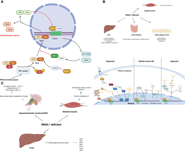
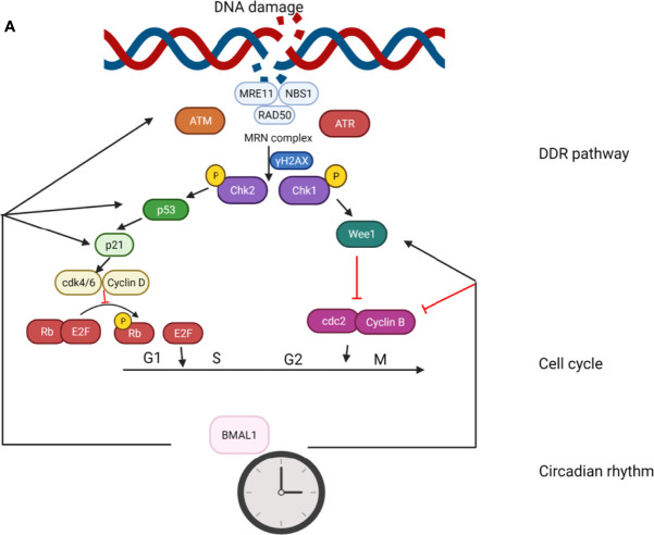
Cell senescence is a crucial process in cell fate determination and is involved in an extensive array of aging-associated diseases. General perceptions and experimental evidence point out that the decline of physical function as well as aging-associated diseases are often initiated by cell senescence and organ ageing. Therefore, regulation of cell senescence process can be a promising way to handle aging-associated diseases such as osteoporosis. The circadian clock regulates a wide range of cellular and physiological activities, and many age-linked degenerative disorders are associated with the dysregulation of clock genes. BMAL1 is a core circadian transcription factor and governs downstream genes by binding to the E-box elements in their promoters. Compelling evidence has proposed the role of BMAL1 in cellular senescence and aging-associated diseases. In this review, we summarize the linkage between BMAL1 and factors of cell senescence including oxidative stress, metabolism, and the genotoxic stress response. Dysregulated and dampened BMAL1 may serve as a potential therapeutic target against aging- associated diseases.
Keywords: BMAL1; aging; cellular senescence; genotoxic stress; metabolism; oxidative stress.
Copyright © 2022 Zhang, Xiong, Tao, Panayi, Mi and Liu.
Conflict of interest statement
The authors declare that the research was conducted in the absence of any commercial or financial relationships that could be construed as a potential conflict of interest.
Figures
Similar articles
-
BMAL1 modulates senescence programming via AP-1.Aging (Albany NY). 2023 Oct 10;15(19):9984-10009. doi: 10.18632/aging.205112. Epub 2023 Oct 10. Aging (Albany NY). 2023. PMID: 37819791 Free PMC article.
-
Circadian clock gene Clock-Bmal1 regulates cellular senescence in Chronic obstructive pulmonary disease.BMC Pulm Med. 2022 Nov 22;22(1):435. doi: 10.1186/s12890-022-02237-y. BMC Pulm Med. 2022. PMID: 36419003 Free PMC article.
-
Bmal1-deficient mouse fibroblast cells do not provide premature cellular senescence in vitro.Chronobiol Int. 2018 May;35(5):730-738. doi: 10.1080/07420528.2018.1430038. Epub 2018 Jan 26. Chronobiol Int. 2018. PMID: 29372841
-
The Circadian Clock, Nutritional Signals and Reproduction: A Close Relationship.Int J Mol Sci. 2023 Jan 12;24(2):1545. doi: 10.3390/ijms24021545. Int J Mol Sci. 2023. PMID: 36675058 Free PMC article. Review.
-
Circadian rhythm, hypoxia, and cellular senescence: From molecular mechanisms to targeted strategies.Eur J Pharmacol. 2025 Mar 5;990:177290. doi: 10.1016/j.ejphar.2025.177290. Epub 2025 Jan 23. Eur J Pharmacol. 2025. PMID: 39863143 Review.
Cited by
-
Circadian-driven tissue specificity is constrained under caloric restricted feeding conditions.Commun Biol. 2024 Jun 20;7(1):752. doi: 10.1038/s42003-024-06421-0. Commun Biol. 2024. PMID: 38902439 Free PMC article.
-
Juvenile zebrafish (Danio rerio) are able to recover from lordosis.Sci Rep. 2022 Dec 13;12(1):21533. doi: 10.1038/s41598-022-26112-2. Sci Rep. 2022. PMID: 36513797 Free PMC article.
-
Disruption of the Clock Component Bmal1 in Mice Promotes Cancer Metastasis through the PAI-1-TGF-β-myoCAF-Dependent Mechanism.Adv Sci (Weinh). 2023 Aug;10(24):e2301505. doi: 10.1002/advs.202301505. Epub 2023 Jun 17. Adv Sci (Weinh). 2023. PMID: 37330661 Free PMC article.
-
Oxidative Stress-Driven Cellular Senescence: Mechanistic Crosstalk and Therapeutic Horizons.Antioxidants (Basel). 2025 Aug 12;14(8):987. doi: 10.3390/antiox14080987. Antioxidants (Basel). 2025. PMID: 40867884 Free PMC article. Review.
-
Identifying novel circadian rhythm biomarkers for diagnosis and prognosis of melanoma by an integrated bioinformatics and machine learning approach.Aging (Albany NY). 2024 Jun 20;16(16):11824-11842. doi: 10.18632/aging.205961. Epub 2024 Jun 20. Aging (Albany NY). 2024. PMID: 39213172 Free PMC article.
References
Publication types
MeSH terms
Substances
LinkOut - more resources
Full Text Sources