. 2022 Jun 6:10:920569.
doi: 10.3389/fcell.2022.920569. eCollection 2022.

HLH-1 Modulates Muscle Proteostasis During Caenorhabditis elegans Larval Development

Affiliations

HLH-1 Modulates Muscle Proteostasis During Caenorhabditis elegans Larval Development

Khairun Nisaa et al. Front Cell Dev Biol. .

Abstract

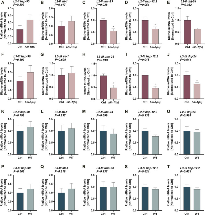
Muscle proteostasis is shaped by the myogenic transcription factor MyoD which regulates the expression of chaperones during muscle differentiation. Whether MyoD can also modulate chaperone expression in terminally differentiated muscle cells remains open. Here we utilized a temperature-sensitive (ts) conditional knockdown nonsense mutation in MyoD ortholog in C. elegans, HLH-1, to ask whether MyoD plays a role in maintaining muscle proteostasis post myogenesis. We showed that hlh-1 is expressed during larval development and that hlh-1 knockdown at the first, second, or third larval stages resulted in severe defects in motility and muscle organization. Motility defects and myofilament organization were rescued when the clearance of hlh-1(ts) mRNA was inhibited, and hlh-1 mRNA levels were restored. Moreover, hlh-1 knockdown modulated the expression of chaperones with putative HLH-1 binding sites in their promoters, supporting HLH-1 role in muscle maintenance during larval development. Finally, mild disruption of hlh-1 expression during development resulted in earlier dysregulation of muscle maintenance and function during adulthood. We propose that the differentiation transcription factor, HLH-1, contributes to muscle maintenance and regulates cell-specific chaperone expression post differentiation. HLH-1 may thus impact muscle proteostasis and potentially the onset and manifestation of sarcopenia.

Keywords: Caenorhabditis elegans (c. elegans); MyoD; chaperone; development; hlh-1; myosin; proteostasis.


Conflict of interest statement

The authors declare that the research was conducted in the absence of any commercial or financial relationships that could be construed as a potential conflict of interest.

Figures

FIGURE 1
FIGURE 1
Differentiation transcription factor HLH-1 is required for muscle maintenance during larval development. (A) Schematic representation of the experimental setup. Wild type (WT) or hlh-1(ts) embryos were placed on seeded plates at 15°C. Animals were maintained at 15°C (Ctrl) or shifted to 25°C at the indicated times, corresponding to specific larval stages (L1-I, L2-II, L3-III, and L4-IV). Young adults (YA, before the onset of egg-laying) were then analyzed for motility, myofilament organization, and chaperone expression. (B) A list of treatments duration (in hours) at 15°C and 25°C for each group (Ctrl and I-IV). (C–D) Motility rates of temperature-shifted YA. Thrashing rates were scored on day one of adulthood for WT (C) or hlh-1(ts) (D) animals grown at 15°C for the duration of the experiment (Ctrl-75 h) or shifted to 25°C at the indicated times (I-15h, II-36h, III-46 h or IV-60 h). Data are means ± 1 standard error of the mean (1SE). Data were analyzed using one-way ANOVA followed by a Dunnett’s post-hoc test (N = 3, n = 30). (*) denotes p < 0.05, (**) denotes p < 0.01, (***) denotes p < 0.001, and (ns) denotes p > 0.05, compared with Ctrl animals maintain at 15°C. (E) Representative images of age-synchronized hlh-1(ts) animals that express MYO-3::GFP. Animals were grown at 15°C for the duration of the experiment (Ctrl) or shifted to 25°C at L1-I or L2-II. Animals were collected and fixed at the YA stage, and myofilaments were imaged. The scale bar is 25 μm. Panels A–B were created using BioRender.com.
FIGURE 2
FIGURE 2
Blocking the RNA-mediated decay pathway rescues animals’ motility. (A–C) Motility rates of wild type (WT), hlh-1(ts), or hlh-1(ts);smg-6. Thrashing rates of age-synchronized WT, hlh-1(ts), or hlh-1(ts);smg-6 grown at 15°C for the experiment duration (A) or shifted to 25°C at L2 [II, (B)] or L3 [III, (C)] larval stages. Data are means ± 1 standard error of the mean (1SE). Data were analyzed using one-way ANOVA followed by a Tukey’s post hoc test (N = 3, n = 20). (*) denotes p < 0.05, and (***) denotes p < 0.001 compared with WT. (D) Representative images of temperature-shifted animals expressing MYO-3:GFP. WT, hlh-1(ts), or hlh-1(ts);smg-6 animals were shifted to 25°C at L1-I or L2-II. YA were collected and fixed, and myofilaments were imaged. The scale bar is 25 μm. Inserts are a 2-fold magnification of the boxed area. (E) Schematic representation of the experimental setup. hlh-1(ts) embryos were laid on seeded plates at 15°C. Age-synchronized hlh-1(ts) animals were moved to RNAi plates seeded with smg-2, smg-7, or empty vector (EV) control, and plates were shifted to 25°C at L1 (I) or L2 (II) larval stages. YAs were then analyzed for motility and myofilament organization. (F–G) Motility rates of hlh-1(ts) smg(RNAi) treated animals. Thrashing rates of age-synchronized hlh-1(ts) animals that were moved to RNAi plates seeded with smg-2, smg-7, or EV control and shifted to 25°C at L1 (F) or L2 (G) larval stages. Data are means ± 1 standard error of the mean (1SE). Data were analyzed using one-way ANOVA followed by a Dunnett’s post hoc test (N = 3, n = 60). (**) denotes p < 0.01 and (***) denotes p < 0.001 compared with EV control. (H) Representative images of hlh-1(ts) smg(RNAi) treated myofilament. Age-synchronized hlh-1(ts) animals expressing MYO-3:GFP were treated with smg-2, smg-7, or EV control RNAi at 25°C as in G, and myofilaments were imaged. The scale bar is 7.5 μm. Panel E was created using BioRender.com.
FIGURE 3
FIGURE 3
The expression of HLH-1-dependent chaperones during larval development. (A–T) Expression levels of chaperones in hlh-1(ts) animals. Relative mRNA levels of HLH-1-dependent chaperones, hsp-90, sti-1, unc-23, hsp-12.2, or dnj-24, in age synchronized hlh-1(ts) (A–J) or WT (K–T) animals, shifted to 25°C at L2-II [(A-E) and (K-O)] or L3-III [(F-J) and (P-T)] larval stages. Expression levels were compared with the same strain animals maintained at 15°C (Ctrl). Data are means ± 1 standard error of the mean (1SE). Data were analyzed using the Wilcoxon Mann-Whitney rank sum test compared with the same strain Ctrl treated animals (N ≥ 4).
FIGURE 4
FIGURE 4
Mild disruption of hlh-1 expression during development severely impact muscle maintenance later in adulthood. (A–C) Motility rates of WT or hlh-1(ts) adults. Thrashing rates of age-synchronized WT or hlh-1(ts) animals maintained at 15°C were examined on days 1 (A), 4 (B), or 7 (C) of adulthood. Data are means ± 1 standard error of the mean (1SE). Data were analyzed using the Wilcoxon Mann-Whitney rank sum test compared with WT (N = 3, n ≥ 30). (D) Representative images of WT or hlh-1(ts) animals that express MYO-3::GFP. Age-synchronized WT or hlh-1(ts) animals maintained at 15°C were collected and fixed on days 1, 4, or 7 of adulthood, and myofilaments were imaged. Inserts are a 2-fold magnification of the boxed area. The scale bar is 10 μm.

Similar articles

Cited by

References

    1. Al-Tahan S., Weiss L., Yu H., Tang S., Saporta M., Vihola A., et al. (2019). New Family with HSPB8-Associated Autosomal Dominant Rimmed Vacuolar Myopathy. Neurol. Genet. 5 (4), e349. 10.1212/NXG.0000000000000349 - DOI - PMC - PubMed
    1. Bar-Lavan Y., Shemesh N., Ben-Zvi A. (2016a). Chaperone Families and Interactions in Metazoa. Essays Biochem. 60 (2), 237–253. 10.1042/EBC20160004 - DOI - PubMed
    1. Bar-Lavan Y., Shemesh N., Dror S., Ofir R., Yeger-Lotem E., Ben-Zvi A. (2016b). A Differentiation Transcription Factor Establishes Muscle-specific Proteostasis in Caenorhabditis elegans . PLoS Genet. 12 (12), e1006531. 10.1371/journal.pgen.1006531 - DOI - PMC - PubMed
    1. Basha O., Argov C. M., Artzy R., Zoabi Y., Hekselman I., Alfandari L., et al. (2020). Differential Network Analysis of Multiple Human Tissue Interactomes Highlights Tissue-Selective Processes and Genetic Disorder Genes. Bioinformatics 36 (9), 2821–2828. 10.1093/bioinformatics/btaa034 - DOI - PubMed
    1. Ben-Zvi A., Miller E. A., Morimoto R. I. (2009). Collapse of Proteostasis Represents an Early Molecular Event in Caenorhabditis elegans Aging. Proc. Natl. Acad. Sci. U.S.A. 106 (35), 14914–14919. 10.1073/pnas.0902882106 - DOI - PMC - PubMed

LinkOut - more resources