Review
. 2022 Jun 3;18(3):39-48.
doi: 10.14797/mdcvj.1093. eCollection 2022.

Imaging: New Frontiers in Vascular Training

Affiliations
Review

Imaging: New Frontiers in Vascular Training

Kavya Sinha et al. Methodist Debakey Cardiovasc J. .

Abstract

Advances in medical imaging have redefined the practice of vascular surgery. Current training programs for vascular surgery do not incorporate formal training in vascular imaging other than in duplex ultrasound when a physician is undergoing the vascular interpretation certification process. Yet imaging modalities and techniques have grown exponentially in the adjacent fields of interventional radiology, interventional and diagnostic cardiology, and neuroradiology, so much so that advanced imaging fellowships have been established in these fields. This article reviews the current state of vascular imaging training, identifies gaps in the current training regimen, and proposes an advanced vascular imaging fellowship for the future.

Keywords: fellowship; medical education; vascular imaging; vascular surgery.


Conflict of interest statement

The authors have no competing interests to declare.

Figures

Figure 1 Integrated 0+5 vascular surgery residency curriculum.
Figure 1
Integrated 0+5 vascular surgery residency curriculum; red boxes indicate training rotations related to imaging.
Figure 2 Tibial vessels imaged with CTA and MRI using T1/T2-weighted and ultrashort echo time sequences.
Figure 2
Tibial vessels imaged with computed tomography angiography (CTA) and magnetic resonance imaging (MRI) using T2-weighted, T1-weighted, and ultrashort echo time sequences. The CTA shows concentric calcium but suffers from beam hardening artifacts and calcium blooming that obscures the vessel lumen. MRI with ultrashort echo time imaging shows a very thin rim of hypointensity around the central vessel, indicating that the calcium ring is thin and the lumen is occluded with mixed morphology plaque.
Figure 3 3D multiplanar reconstruction determines aortic neck measurements to size abdominal aortic endograft.
Figure 3
Three-dimensional multiplanar reconstruction determines aortic neck diameter, angle, and centerline measurements to size abdominal aortic endograft.
Figure 4 Cinematic rendering guides optimal approach for type 4 thoracoabdominal aneurysm repair.
Figure 4
Cinematic rendering helps plan the optimal incision and approach for open repair of type 4 thoracoabdominal aneurysm.
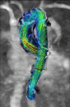
Figure 5 4D flow MRI helps visualize temporal evolution of blood flow patterns within an acquired 3D volume.
Figure 5
An example of four-dimensional (4D) flow magnetic resonance imaging, which enables the visualization of the temporal evolution of complex blood flow patterns within an acquired 3D volume. The blood flow patterns are represented in the colored vectors, which represent velocity with blue being closer to 0 m/s and red up to 1 m/s.
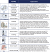
Figure 6 The strengths of an interdisciplinary vascular imaging fellowship team show applications of different imaging modalities.
Figure 6
The strengths and capabilities of an interdisciplinary team in an advanced vascular imaging fellowship demonstrate the various applications of the different imaging modalities. MRI: magnetic resonance imaging; CT: computed tomography; IVUS/OCT: intravascular ultrasound/optical coherence tomography; FFR: fractional flow reserve; CTA: CT angiography; RPVI: Registered Physician in Vascular Interpretation; PAD: peripheral artery disease; TCD: transcranial doppler
Figure 7 The core competencies of our proposed advanced vascular imaging fellowship.
Figure 7
The core competencies of our proposed advanced vascular imaging fellowship. TCD: transcranial doppler; CBCT: cone-beam computed tomography; DCTA: dynamic CT angiography; IVUS: intravascular ultrasound; OCT: optical coherence tomography

References

    1. Smeds MR, Sheahan MG, Shames ML, Abularrage CJ. A modern appraisal of current vascular surgery education. J Vasc Surg. 2021. Apr;73(4):1430-1435. doi: 10.1016/j.jvs.2020.10.028 - DOI - PubMed
    1. Chow BJW, Alenazy A, Small G, et al. Competency-Based Medical Education: Do the Cardiac Imaging Training Guidelines Have it Right? JACC Cardiovasc Imaging. 2019. Dec;12(12):2505-2513. doi: 10.1016/j.jcmg.2019.09.021 - DOI - PubMed
    1. Cheng TW, Farber A, Rajani RR, et al. National criteria for academic appointment in vascular surgery. J Vasc Surg. 2019. May;69(5):1559-1565. doi: 10.1016/j.jvs.2018.08.178 - DOI - PubMed
    1. Halperin JL, Williams ES, Fuster V. COCATS 4 Introduction. J Am Coll Cardiol. 2015. May 5;65(17):1724-33. doi: 10.1016/j.jacc.2015.03.020 - DOI - PubMed
    1. Creager MA, Hamburg NM, Calligaro KD, et al. 2021 ACC/AHA/SVM/ACP Advanced Training Statement on Vascular Medicine (Revision of the 2004 ACC/ACP/SCAI/SVMB/SVS Clinical Competence Statement on Vascular Medicine and Catheter-Based Peripheral Vascular Interventions). Circ Cardiovasc Interv. 2021. Feb;14(2):e000079. doi: 10.1161/HCV.0000000000000079 - DOI - PMC - PubMed