

Macular microcirculation changes after macula-off rhegmatogenous retinal detachment repair with silicone oil tamponade evaluated by OCT-A: preliminary results
- PMID: 35734223
- PMCID: PMC9208039
- DOI: 10.1177/25158414221105222
Macular microcirculation changes after macula-off rhegmatogenous retinal detachment repair with silicone oil tamponade evaluated by OCT-A: preliminary results
Abstract
Background: Rhegmatogenous retinal detachment (RRD) with macular involvement is a sight-threatening condition. Silicone oil (SO) is efficacious for retinal tamponade, especially in complex cases. Whether macular detachment per se or the potential tamponading agent may affect macular microcirculation after RRD repair is a matter of research.
Objectives: To investigate macular microcirculation changes using optical coherence tomography angiography (OCT-A) after pars plana vitrectomy (PPV) with intravitreal SO for RRD repair in the early posttreatment period.
Design: Prospective comparative cross-sectional study.
Data sources and methods: Fourteen eyes of 14 patients were included in the study. All eyes underwent a single successful PPV with SO tamponade for macula-off RRD. OCT-A was performed to analyze macular microcirculation and visual outcomes at 1 month postoperatively. The fellow unaffected eye was used as control.
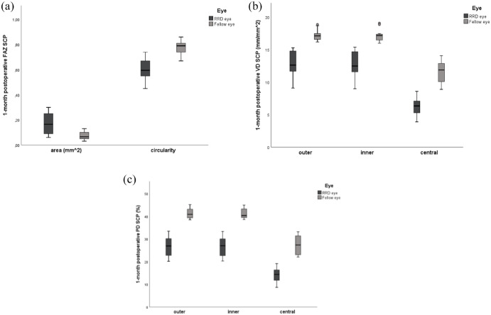
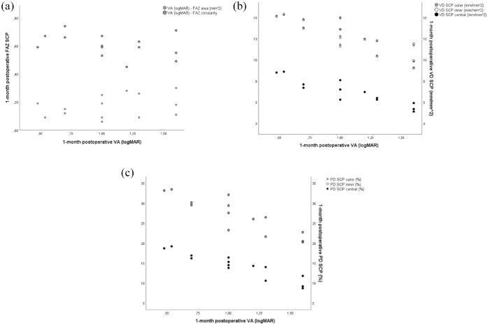
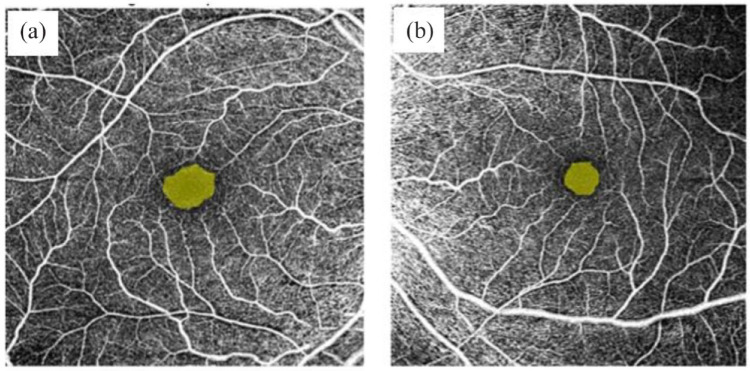
Results: Vessel density (VD) in the superficial capillary plexus (SCP) was significantly lower at each macular region (fovea, parafovea, and perifovea) of SO-treated eyes compared with the fellow eyes (all p = 0.001). Similarly, perfusion density (PD) in the SCP was significantly lower at each macular region than the fellow eyes (all p = 0.001). There was enlargement of foveal avascular zone (FAZ) area and decrease of circularity at RRD eyes compared with the fellow ones (all p = 0.001). Postoperative logMAR visual acuity (VA) was significantly lower in treated eyes than fellow eyes and correlated inversely with foveal, parafoveal, and perifoveal VD and PD SCP (all p < 0.001). Postoperative VA had no correlation with FAZ parameters.
Conclusion: Enlargement of FAZ SCP and decrease in VD and PD SCP during the short-term follow-up were possibly attributable to ischemic changes in the macular area after RRD repair with SO tamponade. In this preliminary study, the flow density in macular capillary plexus may represent an indicator of visual outcomes.
Keywords: macula-off; macular capillary plexus; optical coherence tomography angiography; rhegmatogenous retinal detachment; silicone oil.
© The Author(s), 2022.
Conflict of interest statement
Conflict of interest statement: The authors declared no potential conflicts of interest with respect to the research, authorship, and/or publication of this article.
Figures
Similar articles
-
Effect of long-term silicone oil tamponade on the density of blood vessels in the macular and peripapillary region in patients with rhegmatogenous retinal detachment.Int Ophthalmol. 2025 Mar 25;45(1):124. doi: 10.1007/s10792-025-03460-2. Int Ophthalmol. 2025. PMID: 40131517
-
Evaluation of macular vessel density changes after vitrectomy with silicone oil tamponade in patients with rhegmatogenous retinal detachment.Int J Ophthalmol. 2021 Jun 18;14(6):881-886. doi: 10.18240/ijo.2021.06.14. eCollection 2021. Int J Ophthalmol. 2021. PMID: 34150544 Free PMC article.
-
Microstructural and hemodynamic changes in the fundus after pars plana vitrectomy for different vitreoretinal diseases.Graefes Arch Clin Exp Ophthalmol. 2024 Jul;262(7):1977-1992. doi: 10.1007/s00417-023-06303-x. Epub 2023 Nov 20. Graefes Arch Clin Exp Ophthalmol. 2024. PMID: 37982887 Review.
-
Long-Term Macular Vascular Changes after Primary Rhegmatogenous Retinal Detachment Surgery Resolved with Different Tamponade or Different Surgical Techniques.Life (Basel). 2022 Sep 30;12(10):1525. doi: 10.3390/life12101525. Life (Basel). 2022. PMID: 36294960 Free PMC article.
-
Macular microcirculation changes after repair of rhegmatogenous retinal detachment assessed with optical coherence tomography angiography: A systematic review and meta-analysis.Front Physiol. 2022 Dec 14;13:995353. doi: 10.3389/fphys.2022.995353. eCollection 2022. Front Physiol. 2022. PMID: 36589420 Free PMC article.
Cited by
-
Effect of long-term silicone oil tamponade on the density of blood vessels in the macular and peripapillary region in patients with rhegmatogenous retinal detachment.Int Ophthalmol. 2025 Mar 25;45(1):124. doi: 10.1007/s10792-025-03460-2. Int Ophthalmol. 2025. PMID: 40131517
-
Effect of silicone oil on retinal microcirculation after vitrectomy for rhegmatogenous retinal detachment evaluated by OCT angiography: a literature review.Ther Adv Ophthalmol. 2023 May 24;15:25158414231174145. doi: 10.1177/25158414231174145. eCollection 2023 Jan-Dec. Ther Adv Ophthalmol. 2023. PMID: 37255621 Free PMC article. Review.
-
Functional and perfusion changes associated with silicone oil tamponade after macula-off rhegmatogenous retinal detachment surgery: an optical coherence tomography angiography/microperimetry study.Int Ophthalmol. 2024 Feb 22;44(1):107. doi: 10.1007/s10792-024-03037-5. Int Ophthalmol. 2024. PMID: 38386180 Free PMC article.
References
-
- Kuhn F, Aylward B. Rhegmatogenous retinal detachment: a reappraisal of its pathophysiology and treatment. Ophthalmic Res 2014; 51: 15–31. - PubMed
-
- D’Amico DJ. Clinical practice. Primary retinal detachment. N Engl J Med 2008; 59: 346–354. - PubMed
-
- De la Rúa ER, Pastor JC, Fernández I, et al.. Non-complicated retinal detachment management: variations in 4 years. Br J Ophthalmol 2008; 92: 523–525. - PubMed
LinkOut - more resources
Full Text Sources
Miscellaneous
