

Pediatric brain hydatid cyst about two cases: Case report
- PMID: 35734744
- PMCID: PMC9207044
- DOI: 10.1016/j.amsu.2022.103806
Pediatric brain hydatid cyst about two cases: Case report
Abstract
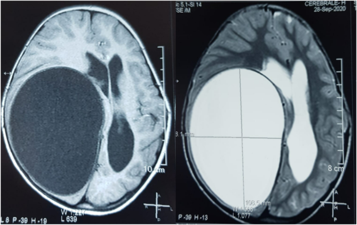
Cerebral hydatid cyst is rare (2%), and mainly affects children. We report 2 cases aged 5 years. The clinical symptomatology was dominated by intracranial hypertension syndrome and motor deficit in both cases. One patient presented a generalized tonic-clonic seizure, the second one presented a left central facial palsy. The diagnosis was made in both cases by brain CT scan and one patient underwent brain MRI. A radiological workup to look for extra-cerebral localization was performed for all patients, which was normal. The treatment was surgical for both patients (D'ARANA-INIGUEZ hydro pulsion technique) with simple after-effects. The postoperative CT scan showed a residual cavity. All our patients were put under antiparasitic treatment with Albendazole.
Keywords: Brain; Case report; Echinococcus; Hydatid cyst; Pediatric.
© 2022 Published by Elsevier Ltd on behalf of IJS Publishing Group Ltd.
Conflict of interest statement
The authors of this article have no conflict or competing interests. All of the authors approved the final version of the manuscript.
Figures


References
-
- Yilmaz N., Kiymaz N., Etlik O., Yazici T. Primary hydatid cyst of the brain during pregnancy. Neurol. Med.-Chir. 2006;46(8):415–417. - PubMed
-
- Agha R.A., Franchi T., Sohrabi C., Mathew G., et al. The SCARE 2020 guideline: updating consensus surgical CAse REport (SCARE) guidelines. Int. J. Surg. 2020;84:226–230. - PubMed
-
- Brahem M., Hlel K., Ayadi A., et al. Cerebral hydatid cysts in children: 4 cases. MedMalInfect. 2006;36(8):434–437. - PubMed
-
- Demir K., Karsli A.F., Kaya T., Devrimci E., Alkan K. Cerebral hydatid cysts: CT findings. Neuroradiology. 1991;33(22–24) - PubMed
Publication types
LinkOut - more resources
Full Text Sources
