Review
. 2022 Sep;190(3):344-357.
doi: 10.1002/ajmg.c.31986. Epub 2022 Jun 23.

C3 glomerulopathy: Understanding an ultra-rare complement-mediated renal disease

Affiliations
Review

C3 glomerulopathy: Understanding an ultra-rare complement-mediated renal disease

Amanda K Heiderscheit et al. Am J Med Genet C Semin Med Genet. 2022 Sep.

Abstract

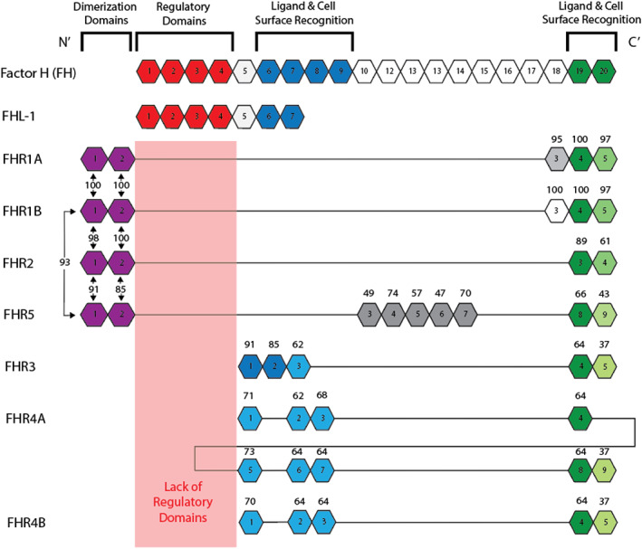
C3 glomerulopathy (C3G) describes a pathologic pattern of injury diagnosed by renal biopsy. It is characterized by the dominant deposition of the third component of complement (C3) in the renal glomerulus as resolved by immunofluorescence microscopy. The underlying pathophysiology is driven by dysregulation of the alternative pathway of complement in the fluid-phase and in the glomerular microenvironment. Characterization of clinical features and a targeted evaluation for indices and drivers of complement dysregulation are necessary for optimal patient care. Autoantibodies to the C3 and C5 convertases of complement are the most commonly detected drivers of complement dysregulation, although genetic mutations in complement genes can also be found. Approximately half of patients progress to end-stage renal disease within 10 years of diagnosis, and, while transplantation is a viable option, there is high risk for disease recurrence and allograft failure. This poor outcome reflects the lack of disease-specific therapy for C3G, relegating patients to symptomatic treatment to minimize proteinuria and suppress renal inflammation. Fortunately, the future is bright as several anti-complement drugs are currently in clinical trials.

Keywords: C3 glomerulonephritis; C3 glomerulopathy; dense deposit disease.


Conflict of interest statement

The authors declare no potential conflict of interest.

Figures

FIGURE 1
FIGURE 1
The complement cascade in C3 glomerulopathy. The complement pathway is activated through three pathways—the classical (CP), lectin (LP), and alternative (AP). The CP is induced by the complement component 1 (C1) complex binding to antibody complexes, pathogen‐associated molecular patterns (PAMPs), or damage‐associated molecular patterns (DAMPs), whereas the LP recognizes microbial polysaccharides. Upon activation, complement component 4 (C4) and complement component 2 (C2) are cleaved by proteases to form C4bC2a, the C3 convertase of the CP and LP. The AP is activated by the spontaneous hydrolysis of complement component 3 (C3) to form C3(H2O), which interacts with factor B and factor D to form C3(H2O)Bb, the initial C3 convertase of the AP. C3 convertases of the CP, LP, and AP cleave C3 into C3a and C3b. C3b binds with factor B and is cleaved by factor D to form C3bBb, the major C3 convertase of the AP. C3bBb cleaves additional C3–C3b and C3a amplifying the complement response. As local levels of C3b increase, C3b associates with C3bBb to form C3bBbC3b, the C5 convertase of the AP. C5 convertase cleaves C5–C5a and C5b. C5b associates with complement components 6–8 (C6–8) and complement component 9 (C9) to from membrane attack complex, which lyses cells, or soluble C5b–9, which is cleared from circulation. C3 glomerulopathy is caused by dysregulation of the AP, although a small percentage of cases are triggered by the CP and LP. Regulators of complement are shown in red, and the sole activator of the complement system, properdin, is shown in green
FIGURE 2
FIGURE 2
Regulators of complement in the glomerular microenvironment. Fluid phase and membrane‐bound regulators play a pivotal role in protection and selectivity of complement activity in the glomerular microenvironment. CR1 inhibitor and C4BP are fluid phase regulators of the CP and LP. Fluid phase regulators of the AP include FH, FHL‐1, and FI. The functions of FH include destabilization of C3bBb by decay accelerating activity, inhibition of FB binding to C3b, and cofactor activity with FI to inactivate C3b to iC3b. The FH splice variant, FHL‐1, also has decay accelerating activity and cofactor activity. DAF, MCP, and CR1 are membrane‐bound regulators of the AP that destabilize C3bBb through cofactor activity and/or decay accelerating activity. Additionally, thrombomodulin is a membrane‐bound regulator that uniquely enhances FH cofactor activity for C3 convertase dissociation. CD59, vitronectin (S protein) and clusterin inhibit the terminal pathway by preventing MAC formation. Thrombomodulin, MCP and CR1 are transmembrane receptors while CD59 and DAF are GPI‐anchored receptors. AP, alternative pathway; C1, complement component 1; C3, complement component 3; C4BP, complement component 4 binding protein; CP, classical pathway; CR1, Complement receptor 1; DAF, decay accelerating factor; FB, factor B; FH, factor H; FHL‐1, factor H‐like 1; FI, factor I; GPI, glycosylphosphatidylinositol; LP, lectin pathway; MAC, membrane attack complex; MASP1‐2, mannose binding ligand associated serine proteases 1–2; MCP, membrane cofactor protein
FIGURE 3
FIGURE 3
The Factor H protein family in humans. All members of the factor H (FH) protein family are built on modules of ~60 amino acids called SCRs that have specific functions. FH has 20 SCRs. The four N′‐terminal SCRs (SCRs 1–4) mediate complement regulation, while SCRs 6–9 and the C′‐terminal SCRs 19–20 recognize ligands in the glycocalyx such as heparan sulfate (HS). Factor H like 1 (FHL‐1) is a splice variant of FH that shares N′‐terminal regulatory functions with FH. SCRs 6–7 of FHL‐1 also recognize ligand and cell surfaces like FH SCRs 6–8. In comparison, factor H‐related proteins (FHRs) 1, 2, and 5 (called type 1 FHRs) lack the N′‐terminal regulatory domains of FH but do have similar C′‐terminal SCRs. Like SCRs 19–20 of FH, the last two domains of type 1 FHRs recognize cell surface ligands such as HS. The N′‐terminal SCRs 1–2 of the type 1 FHRs are essential for homo‐ and hetero‐dimerization. Type 2 FHRs (FHR3 and FHR4) lack regulatory domains but share sequence similarity with the last two SCRs of FH. As a consequence of this sequence similarity, FH and FHRs functionally compete in the local glomerular microenvironment. (In this figure, SCRs are represented by hexagons and aligned by sequence similarity. Amino acid similarities are indicated by number percentages relative to FH or to dimerization domains of type 1 FHRs. Binding and activation sites are indicated by color. A and B for FHR1 indicate acidic and basic forms. A and B for FHR4 indicate long and short isoforms)
FIGURE 4
FIGURE 4
Complement in the glycocalyx. Complement effects on C3 glomerulopathy (C3G) are most prominent in the glomerular microenvironment. Within a kidney nephron, the glomerulus is the filtration unit and is made up of several filtration layers, including the glycocalyx, endothelial cells, glomerular basement membrane (GBM), and podocytes. Together, they retain valuable proteins and filter solutes/waste products into the Bowman's space to be excreted as urine. The glycocalyx, the first filtration barrier, is a highly charged layer of membrane‐bound macromolecules attached to glomerular endothelial cell and the GBM overlying the glomerular endothelial pores, which are termed fenestrae. The glycocalyx is composed of glycosaminoglycans and proteoglycans linked to the cytoskeleton to provide stability as well as cell signaling. Important glycosaminoglycans to note are heparan sulfate (HS), chondroitin sulfate (CS), and nonsulfated hyaluronan (Martinez‐Seara Monne, Danne, Róg, Ilpo, & Gurtovenko, 2013). Nonsulfated hyaluronan forms a stiff, random coil that allows small molecules to diffuse freely while excluding larger molecules. HS and CS are distributed on all cell surfaces and in extracellular matrices and bind a variety of ligands (Izumikawa & Kitagawa, 2010). HS in particular binds factor H (FH) and factor H‐related proteins (FHRs; Blackmore et al., ; Clark et al., ; Skerka, Chen, Fremeaux‐Bacchi, & Roumenina, 2013). FH binding regulates formation of C3 convertase while FHRs promote C3 convertase activation. The local balance between FH and FHRs impacts complement control and when altered can potentiate glomerular disease (Csincsi et al., ; Goodship et al., ; Loeven et al., ; Tortajada et al., 2013). (a) The balance between FH and FHRs favors complement control. (b) Increased binding of FHRs (FHR1 is shown) and decreased binding of FH drives complement dysregulation
FIGURE 5
FIGURE 5
Differential diagnosis of C3 dominant glomerulonephritis. Complement component 3 (C3) dominant staining is defined as C3 staining greater by at least two orders of magnitude more than any other immunoreactant in renal tissue. Disease processes that fall into this category are monoclonal gammopathy of renal significance (MGRS), post‐infectious glomerulonephritis (PIGN), and C3 glomerulopathy (C3G). If C3 dominant glomerulonephritis is diagnosed, patients >50 years of age should be screed for paraproteins. During screening, protease digestion prior to IF can unmask immunoglobulins (Igs). This treatment will help avoid misdiagnosis of C3G, as shown by recent studies demonstrating false‐negative staining of Igs on routine IF (Larsen et al., 2015). Ig staining equal to C3 staining at first biopsy; however, does not rule out genetic or acquired drivers of AP dysregulation. C3 dominant staining is also observed in ~30% of PIGN, which resembles C3G on renal biopsy making it necessary to monitor C3 serum levels for normalization. If C3 levels normalize after ~8 weeks PIGN is the likely diagnosis; if C3 levels remains abnormal for 12 weeks or greater, a biopsy is warranted and high on the differential diagnosis is C3G. C3G is a pathological description of a disease process that includes two subgroups defined by differences on electron microscopy (EM)–C3 glomerulonephritis (C3GN) and dense deposits disease (DDD). C3GN accounts for ~66% of C3G cases and on EM has mesangial, subepithelial, subendothelial, and/or less dense, discontinuous, ill‐defined intramembranous deposits. DDD accounts for ~33% of C3G cases and on EM has findings of sausage‐shaped, electron dense intramembranous deposits that thicken the lamina densa. Light microscopy cannot differentiate C3G from other glomerulonephritis
FIGURE 6
FIGURE 6
Biomarker distribution of C3 and soluble C5b‐9 in C3 glomerulopathy. (a) Of 285 C3 glomerulopathy (C3G) patients, 42% had low complement component 3 (C3) serum levels providing fluid‐phase evidence of increased complement activity at the level of the C3 convertase, however in 58% serum C3 was normal. (b) Of 416 C3G patients, 56% had increased soluble C5b‐9 (sC5b‐9) levels, a fluid‐phase indication of terminal pathway activity; in 44%, sC5b‐9 was normal. (c) In aggregate, of 280 patients with both sC5b‐9 and C3 testing, 172 (61.4%) had fluid‐phase evidence of complement dysregulation, while in 108 patients (38.6%), circulating levels of complement components were normal
FIGURE 7
FIGURE 7
Treatment algorithm for C3 glomerulopathy. Patients who present with proteinuria, hematuria (macroscopic or microscopic), hypertension, or signs and symptoms of renal function impairment (renal insufficiency, nephrotic syndrome, and/or nephritic syndrome) should have a complete blood count, complement component 3 (C3), complement component 4 (C4), antinuclear antibodies, antineutrophil cytoplasmic antibodies, and urine protein creatinine, as well as screening for hepatitis and other infections if indicated. Persistent proteinuria, a reduce glomerular filtration rate, and/or unexplained hypertension are considerations for kidney biopsy. Based on biopsy findings, current classification separates patients into IC‐MPGN with significant glomerular Ig and complement deposition, and C3G with characteristic C3 dominant glomerulonephritis; however, recent evidence suggests some overlap between C3G and IC‐MPGN. In patients with biopsy confirmed C3 dominant glomerulonephritis, PIGN and MGRS should be ruled out. If C3 glomerulopathy is diagnosed, a battery of complement testing for biomarkers, acquired drivers, functional assays, and genetic abnormalities is indicated. Results help to create an individualized treatment plan. Angiotensin‐converting enzyme (ACE) inhibitors and/or ARBs are first line therapy for proteinuria and reduce blood pressure. Persistent proteinuria warrants a trial with mycophenolate mofetil (MMF) and if needed, a short course of steroids. If renal function continues to deteriorate, clinicians should encourage patients to consider the many ongoing clinical trials evaluating novel anti‐complement therapies (see ClinicalTrials.gov). If terminal pathway activity is identified, eculizumab can be considered

References

    1. Allison, A. C. (2005). Mechanisms of action of mycophenolate mofetil. Lupus, 14(3_suppl), 2–8. 10.1191/0961203305lu2109oa - DOI - PubMed
    1. Angelo, J. R. , Bell, C. S. , & Braun, M. C. (2011). Allograft failure in kidney transplant recipients with membranoproliferative glomerulonephritis. American Journal of Kidney Diseases, 57(2), 291–299. 10.1053/j.ajkd.2010.09.021 - DOI - PubMed
    1. Avasare, R. S. , Canetta, P. A. , Bomback, A. S. , Marasa, M. , Caliskan, Y. , Ozluk, Y. , … Appel, G. B. (2018). Mycophenolate mofetil in combination with steroids for treatment of C3 glomerulopathy: A case series. Clinical Journal of the American Society of Nephrology, 13(3), 406–413. 10.2215/CJN.09080817 - DOI - PMC - PubMed
    1. Banks, R. , May, S. , & Willington, T. (1982). Acute renal failure in dense deposit disease: Recovery after plasmapheresis. British Medical Journal (Clinical Research Ed.), 284(6332), 1874–1875. - PMC - PubMed
    1. Blackmore, T. K. , Hellwage, J. , Sadlon, T. A. , Higgs, N. , Zipfel, P. F. , Ward, H. M. , & Gordon, D. L. (1998). Identification of the second heparin‐binding domain in human complement factor H. Journal of Immunology, 160(7), 3342–3348. - PubMed

Publication types

MeSH terms