. 2022 May 29;12(6):166.
doi: 10.3390/bs12060166.

The Impact of Chronic Unpredictable Mild Stress-Induced Depression on Spatial, Recognition and Reference Memory Tasks in Mice: Behavioral and Histological Study

Affiliations

The Impact of Chronic Unpredictable Mild Stress-Induced Depression on Spatial, Recognition and Reference Memory Tasks in Mice: Behavioral and Histological Study

Ghofran Khalid Alqurashi et al. Behav Sci (Basel). .

Abstract

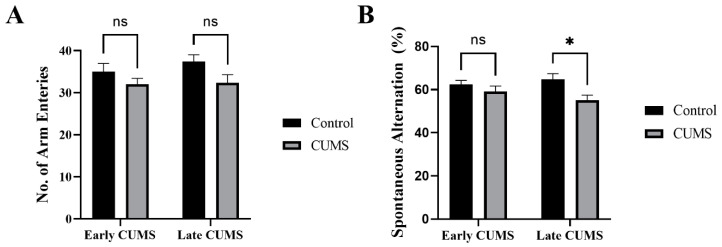
Depression-induced cognitive impairment has recently been given more attention in research. However, the relationship between depression and different types of memory is still not clear. Chronic unpredictable mild stress (CUMS) is a commonly used animal model of depression in which animals are exposed to chronic unpredictable environmental and psychological stressors, which mimics daily human life stressors. This study investigated the impact of different durations of CUMS on various types of memory (short- and long-term spatial memory and recognition memory) and investigated CUMS' impact on the ultrastructural level by histological assessment of the hippocampus and prefrontal cortex. Twenty male C57BL/J6 mice (6 weeks old, 21.8 ± 2 g) were randomly divided into two groups (n = 10): control and CUMS (8 weeks). A series of behavioral tasks were conducted twice at weeks 5-6 (early CUMS) and weeks 7-8 (late CUMS). A tail-suspension test (TST), forced swimming test (FST), elevated zero maze (EZM), elevated plus maze (EPM), open field test (OFT), and sucrose-preference test (SPT) were used to assess anxiety and depressive symptoms. The cognitive function was assessed by the novel object recognition test (NORT; for recognition memory), Y-maze (for short-term spatial memory), and Morris water maze (MWM: for long-term spatial memory) with a probe test (for reference memory). Our data showed that 8 weeks of CUMS increased the anxiety level, reported by a significant increase in anxiety index in both EPM and EZM and a significant decrease in central preference in OFT, and depression was reported by a significant increase in immobility in the TST and FST and sucrose preference in the SPT. Investigating the impact of CUMS on various types of memory, we found that reference memory is the first memory to be affected in early CUMS. In late CUMS, all types of memory were impaired, and this was consistent with the abnormal histological features of the memory-related areas in the brain (hippocampus and prefrontal cortex).

Keywords: anxiety; depression; recognition memory; spatial memory; stress.


Conflict of interest statement

The authors declare no conflict of interest.

Figures

Figure 1
Figure 1
(A) Timeline of the experiment with an overview of behavioral tasks. The same behavioral tests were repeated in weeks 5–6 (early CUMS) and 7–8 (late CUMS). (B) Schematic representation of OFT under EthoVision tracking system. (C) SPT protocol. (D) NORT protocol includes 3 stages: habituation, familiarization, and test stages. (E) MWM protocol. CUMS: chronic unpredictable mild stress, EPM: elevated plus maze, EZM: elevated zero maze, TST: tail suspension test, FST: forced swimming test, OFT: open field test, SPT: sucrose-preference test, NORT: novel-object-recognition test, MWM: Morris water maze, N: north, E: east, SE: southeast, NW: northwest, SW: southwest, NE: northeast. Created with BioRender.com (accessed on 1 November 2021).
Figure 2
Figure 2
The effect of CUMS on the weekly percentage of weight change. Data are presented as mean ± standard error of the mean (SEM). Two-way repeated-measures ANOVA was used, followed by Šídák’s multiple-comparisons test. (*) indicates a significant difference between the CUMS and the control group at p > 0.05 and ** p < 0.01.
Figure 3
Figure 3
The effect of early and late CUMS on locomotor activity in the OFT. (A) Velocity (cm/s); (B) total distance moved (cm); (C) immobility frequency. Two-way ANOVA was used, followed by Šídák’s multiple-comparisons test. OFT: open field test; ns: not significant.
Figure 4
Figure 4
The effect of early and late CUMS on anxiety in (A) central preference percentage, (B) EZM, and (C) EPM. Two-way ANOVA was used, followed by Šídák’s multiple-comparisons test. (**) indicates a significant difference between the CUMS groups and the control group at p < 0.01, *** p < 0.001, and **** p < 0.0001. ns: not significant.
Figure 5
Figure 5
The effect of early and late CUMS on depression tests in the (A) TST, (B) FST, and (C) SPT. Two-way ANOVA was used, followed by Šídák’s multiple-comparisons test. (*) indicates a significant difference between the CUMS groups and the control group at p > 0.05, ** p < 0.01. ns: not significant.
Figure 6
Figure 6
The effect of early and late CUMS on short-term spatial memory in the Y-maze task: (A) number of arm entries and (B) spontaneous alternation. Two-way ANOVA was used, followed by Šídák’s multiple-comparisons test. (*) indicates a significant difference between the treated CUMS and the control group at p > 0.05. ns: not significant.
Figure 7
Figure 7
The effect of CUMS on long-term spatial memory in the MWM task. The effect of early (A) and late (B) CUMS on escape latency time and (C) probe test. Two-way ANOVA was used, followed by Šídák’s multiple-comparisons test. (*) indicates a significant difference between the CUMS and the control group at p > 0.05, *** p < 0.001, and **** p < 0.0001.
Figure 8
Figure 8
The effect of CUMS on recognition memory in the NORT task. The frequency of sniffing familiar objects (F1 and F2) in the familiarization stage in early (A) and late (B) CUMS. The frequency of sniffing objects (F1 and novel) in the test stage in early (C) and late (D) CUMS. The discrimination index (DI) is shown in (E). Two-way ANOVA was used, followed by Šídák’s multiple-comparisons test. (**) indicates a significant difference between the CUMS and the control group at p < 0.01, *** p < 0.001, and **** p < 0.0001. ns: not significant.
Figure 9
Figure 9
The hippocampus structure. H&E staining of hippocampus area sagittal section in both controls (A) and CUMS (B). The hippocampus areas CA1, CA3, and the dentate gyrus (DG) are the main areas that are involved in memory function. There is no interstitial bleeding or infiltration of inflammatory cells in CUMS group. Scale bar = 200 µm.
Figure 10
Figure 10
The effect of CUMS on the hippocampal regions. Representative images of hippocampus areas (CA1 and CA3) and dentate gyrus (DG) from control (A,C,E) and CUMS (B,D,F). The controls had normal neurons with vesicular pale nuclei in CA1 (green arrow) (A) while the control CA3 showed normal large basophilic neurons with dark vesicular nuclei and clear axons (green arrow) (C). However, both CA1 and CA3 in CUMS (B,D, respectively) had degenerated neurons (black arrow) and loss of tissue surrounding the degenerated neurons (red arrow). A dark eosinophilic plaque (blue arrow) was observed in CUMS CA3 (D). The dentate gyrus (DG) polymorphic layer (PL) is thinner in CUMS (E) compared with controls (F). There was no significant change in the DG granular layer (GL) in both groups. Scale bar = 20 µm.
Figure 11
Figure 11
The effect of CUMS on the frontal cortex. Representative frontal cortex areas stained with H&E from controls (A,C) and CUMS (B,D). The control frontal cortex showed the pia matter (brown arrow) and 6 cortical layers (from I to VI) and part of the corpus callosum (CC) in low-power images (A). The cortical layers disrupted by abnormal cellular overgrowth area (Ab) in CUMS frontal cortex (B). The high-power image observed normal neurons with large vesicular nuclei and clear nucleolus and chromatin materials in the control frontal cortex (green arrow) (C). The cellular overgrowth area in CUMS cortex contains degenerated neurons (black arrow), eosinophilic extracellular plaques (blue arrow), and neurons with dark basophilic nuclei without clear nucleolus. (D). Scale bar A,B = 200 µm, C,D = 20 µm.

References

    1. Melchior M., Caspi A., Milne B.J., Danese A., Poulton R., Moffitt T.E. Work stress precipitates depression and anxiety in young, working women and men. Psychol. Med. 2007;37:1119–1129. doi: 10.1017/S0033291707000414. - DOI - PMC - PubMed
    1. Tolentino J.C., Schmidt S.L. DSM-5 Criteria and Depression Severity: Implications for Clinical Practice. Front. Psychiatry. 2018;9:450. doi: 10.3389/fpsyt.2018.00450. - DOI - PMC - PubMed
    1. Kessler R.C., Bromet E.J. The Epidemiology of Depression across Cultures. Annu. Rev. Public Health. 2013;34:119–138. doi: 10.1146/annurev-publhealth-031912-114409. - DOI - PMC - PubMed
    1. Verbeek E.C., Bevova M.R., Hoogendijk W.J.G., Heutink P. The genetics of MDD—A review of challenges and opportunities. J. Depress. Anxiety. 2014;3:150.
    1. Bromet E., Andrade L.H., Hwang I., Sampson N.A., Alonso J., De Girolamo G., De Graaf R., Demyttenaere K., Hu C., Iwata N., et al. Cross-national epidemiology of DSM-IV major depressive episode. BMC Med. 2011;9:90. doi: 10.1186/1741-7015-9-90. - DOI - PMC - PubMed

LinkOut - more resources