. 2022 May 25;9(6):168.
doi: 10.3390/jcdd9060168.

The Time-Dependent Role of Bisphosphonates on Atherosclerotic Plaque Calcification

Affiliations

The Time-Dependent Role of Bisphosphonates on Atherosclerotic Plaque Calcification

Amirala Bakhshian Nik et al. J Cardiovasc Dev Dis. .

Abstract

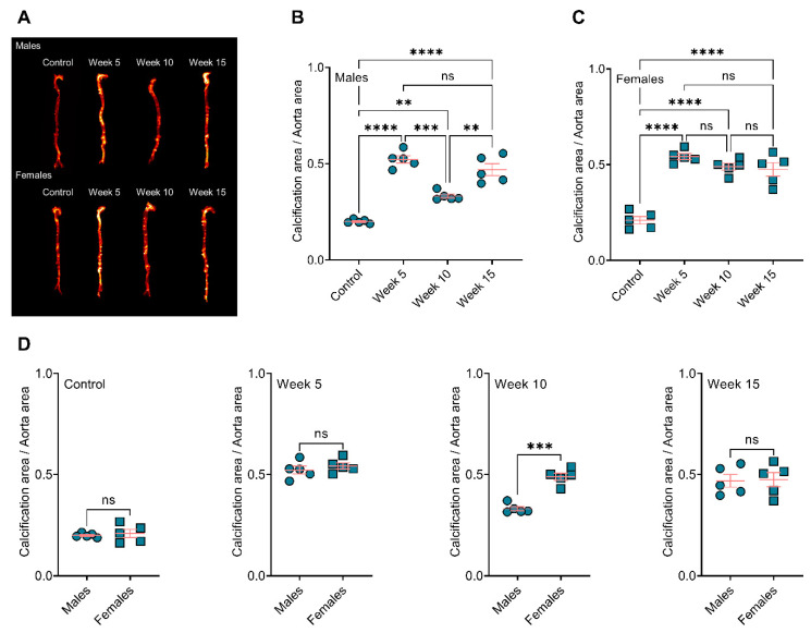
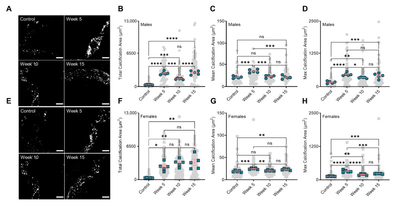
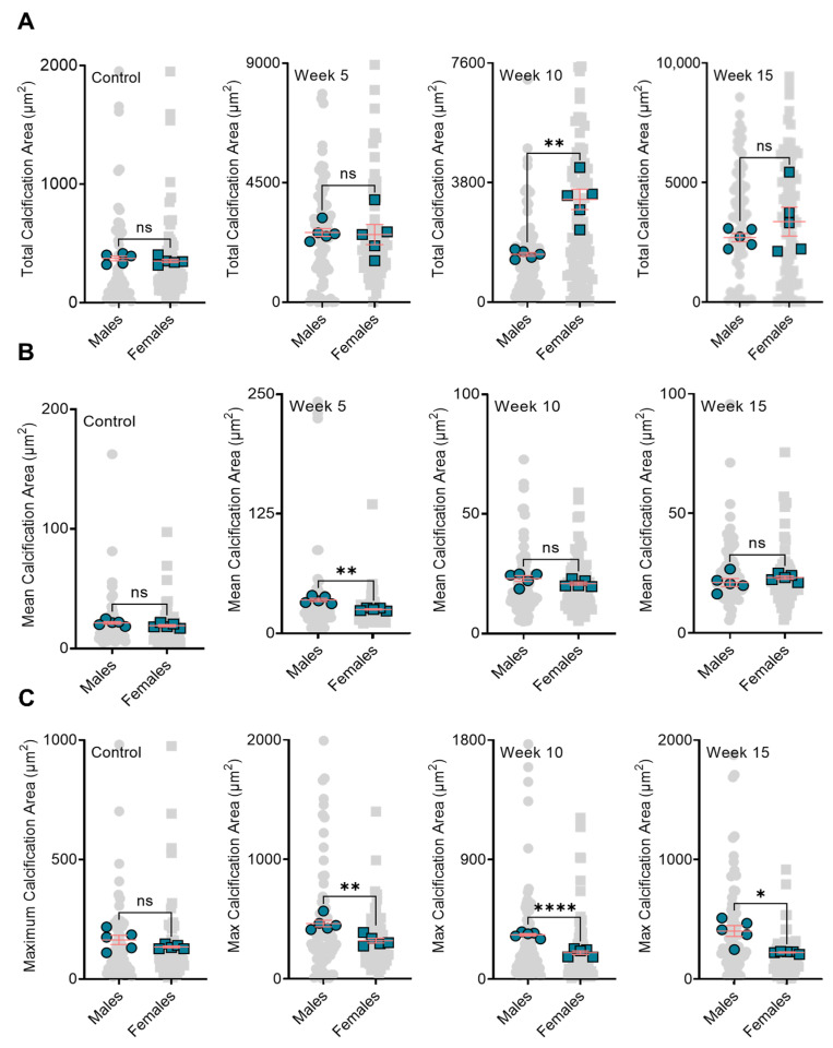
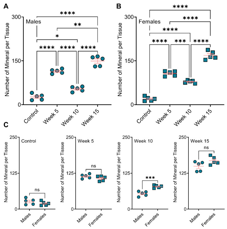
Atherosclerotic plaque calcification directly contributes to the leading cause of morbidity and mortality by affecting plaque vulnerability and rupture risk. Small microcalcifications can increase plaque stress and promote rupture, whereas large calcifications can stabilize plaques. Drugs that target bone mineralization may lead to unintended consequences on ectopic plaque calcification and cardiovascular outcomes. Bisphosphonates, common anti-osteoporotic agents, have elicited unexpected cardiovascular events in clinical trials. Here, we investigated the role of bisphosphonate treatment and timing on the disruption or promotion of vascular calcification and bone minerals in a mouse model of atherosclerosis. We started the bisphosphonate treatment either before plaque formation, at early plaque formation times associated with the onset of calcification, or at late stages of plaque development. Our data indicated that long-term bisphosphonate treatment (beginning prior to plaque development) leads to higher levels of plaque calcification, with a narrower mineral size distribution. When given later in plaque development, we measured a wider distribution of mineral size. These morphological alterations might be associated with a higher risk of plaque rupture by creating stress foci. Yet, bone mineral density positively correlated with the duration of the bisphosphonate treatment.

Keywords: atherosclerotic plaque calcification; bisphosphonates; calcification paradox.


Conflict of interest statement

The authors declare no conflict of interest.

Figures

Figure 1
Figure 1
BiP treatment increases the atherosclerotic plaque calcification in ApoE−/− mice fed an atherogenic diet. (A) Visualization of the calcium burden using a near-infrared calcium tracer, OsteoSense; (B,C) quantification of the OsteoSense signal and correlation to total calcification in male and female mice, respectively; (D) comparison of total calcification between male and female mice in each group. ** p ≤ 0.01, *** p ≤ 0.001, and **** p ≤ 0.0001, two-way ANOVA with Tukey’s post-hoc test was used for comparison between multiple groups, and Student’s t-test was used for comparison between two groups. “ns” stands for not significant.
Figure 2
Figure 2
BiP treatment alters the micromorphology of the minerals in the atherosclerotic plaque. (A) Visualization of the minerals in the atherosclerotic plaques of male mice (20×, scale bar 100 µm); (B) total plaque calcification in male mice; (C) mean calcification area (mean of mineral size) in male animals; (D) maximum calcification area in male mice; (E) visualization of the minerals in the atherosclerotic plaques of female mice (20×, scale bar 100 µm); (F) total plaque calcification in female mice; (G) mean calcification area (mean of mineral size) in female animals; (H) maximum calcification area in female mice. Note that gray points (● or ■) in the background represent technical replications, i.e., all calcifications measured across all mice, and green points (● or ■) represent the biological replications, i.e., the average value for each mouse. * p < 0.05, ** p ≤ 0.01, *** p ≤ 0.001, and **** p ≤ 0.0001, two-way ANOVA with Tukey’s post-hoc test. “ns” stands for not significant.
Figure 3
Figure 3
BiP treatment may affect the mineral morphology differently in male and female mouse model. Panel (A) is the comparison of total plaque calcification between male and female mice; Panel (B) is the comparison of mean calcification area between male and female mice; Panel (C) is the comparison of maximum calcification area between male and female mice. Note that gray points (● or ■) in the background represent technical replications, i.e., all calcifications measured across all mice, and green points (● or ■) represent the biological replications, i.e., the average value for each mouse. * p < 0.05, ** p ≤ 0.01, and **** p ≤ 0.0001, Student’s t-test. “ns” stands for not significant.
Figure 4
Figure 4
BiP treatment increases the number of minerals in the atherosclerotic plaque of ApoE−/− mice. Number of the mineral per tissue in (A) male and (B) female mice; (C) comparison of total plaque calcification between male and female mice. * p < 0.05, ** p ≤ 0.01, *** p ≤ 0.001 and **** p ≤ 0.0001, two-way ANOVA with Tukey’s post-hoc test was used for comparison between multiple groups, and Student’s t-test was used for comparison between two groups. “ns” stands for not significant.
Figure 5
Figure 5
BiP treatment did not affect the serum TNAP activity and total cholesterol in mouse model of atherosclerotic plaque calcification. Serum TNAP activity in (A) male mice and (B) female mice; serum total cholesterol in (C) male mice and (D) female mice. No statistically significance observed across the groups, two-way ANOVA with Tukey’s post-hoc test.
Figure 6
Figure 6
Bone remodeling positively correlated with duration of BiP treatment. (A) Reconstruction of femoral bone microstructure for the different treated and untreated groups; Panel (B) is the bone volume fraction (bone volume/total volume) for the cortical and trabecular regions; Panel (C) is the bone thickness for the cortical and trabecular regions. * p < 0.05, ** p ≤ 0.01, *** p ≤ 0.001, and **** p ≤ 0.0001, two-way ANOVA with Tukey’s post-hoc test. Male mice (●) and female mice (■). “ns” stands for not significant.

References

    1. Ruiz J.L., Hutcheson J.D., Cardoso L., Bakhshian Nik A., Condado de Abreu A., Pham T., Buffolo F., Busatto S., Federici S., Ridolfi A., et al. Nanoanalytical analysis of bisphosphonate-driven alterations of microcalcifications using a 3D hydrogel system and in vivo mouse model. Proc. Natl. Acad. Sci. USA. 2021;118:e1811725118. doi: 10.1073/pnas.1811725118. - DOI - PMC - PubMed
    1. Hutcheson J.D., Goettsch C., Bertazzo S., Maldonado N., Ruiz J.L., Goh W., Yabusaki K., Faits T., Bouten C., Franck G., et al. Genesis and growth of extracellular-vesicle-derived microcalcification in atherosclerotic plaques. Nat. Mater. 2016;15:335–343. doi: 10.1038/nmat4519. - DOI - PMC - PubMed
    1. Bentzon J.F., Otsuka F., Virmani R., Falk E. Mechanisms of Plaque Formation and Rupture. Circ. Res. 2014;114:1852–1866. doi: 10.1161/CIRCRESAHA.114.302721. - DOI - PubMed
    1. Virani S.S., Alonso A., Aparicio H.J., Benjamin E.J., Bittencourt M.S., Callaway C.W., Carson A.P., Chamberlain A.M., Cheng S., Delling F.N., et al. Heart Disease and Stroke Statistics—2021 Update. Circulation. 2021;143:e254–e743. doi: 10.1161/CIR.0000000000000950. - DOI - PubMed
    1. Peng A.W., Mirbolouk M., Orimoloye O.A., Osei A.D., Dardari Z., Dzaye O., Budoff M.J., Shaw L., Miedema M.D., Rumberger J., et al. Long-Term All-Cause and Cause-Specific Mortality in Asymptomatic Patients with CAC ≥ 1000. JACC Cardiovasc. Imaging. 2020;13:83–93. doi: 10.1016/j.jcmg.2019.02.005. - DOI - PMC - PubMed

LinkOut - more resources