. 2022 May 25;8(6):151.
doi: 10.3390/jimaging8060151.

Bladder Wall Segmentation and Characterization on MR Images: Computer-Aided Spina Bifida Diagnosis

Affiliations

Bladder Wall Segmentation and Characterization on MR Images: Computer-Aided Spina Bifida Diagnosis

Rania Trigui et al. J Imaging. .

Abstract

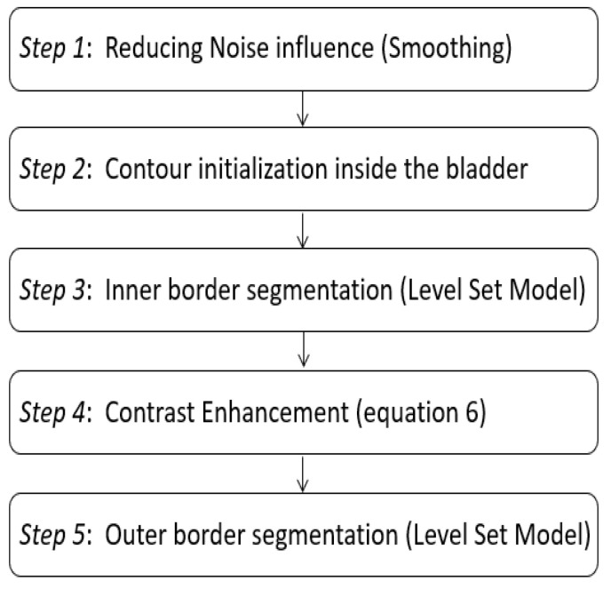
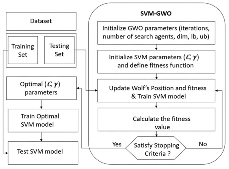
(1) Background: Segmentation of the bladder inner's wall and outer boundaries on Magnetic Resonance Images (MRI) is a crucial step for the diagnosis and the characterization of the bladder state and function. This paper proposes an optimized system for the segmentation and the classification of the bladder wall. (2) Methods: For each image of our data set, the region of interest corresponding to the bladder wall was extracted using LevelSet contour-based segmentation. Several features were computed from the extracted wall on T2 MRI images. After an automatic selection of the sub-vector containing most discriminant features, two supervised learning algorithms were tested using a bio-inspired optimization algorithm. (3) Results: The proposed system based on the improved LevelSet algorithm proved its efficiency in bladder wall segmentation. Experiments also showed that Support Vector Machine (SVM) classifier, optimized by Gray Wolf Optimizer (GWO) and using Radial Basis Function (RBF) kernel outperforms the Random Forest classification algorithm with a set of selected features. (4) Conclusions: A computer-aided optimized system based on segmentation and characterization, of bladder wall on MRI images for classification purposes is proposed. It can significantly be helpful for radiologists as a part of spina bifida study.

Keywords: bladder wall segmentation; classification; magnetic resonance imaging; optimization; sequential floating selection; texture analysis.


Conflict of interest statement

The authors declare no conflict of interest.

Figures

Figure 6
Figure 6
Segmentation procedure using our proposed algorithms: (a) Contour initialization inside the bladde, (b) the inner boundary segmented by the level set algorithm, (c) contrast enhanced generated image, (d) pixel intensity change inside the bladder according to the average intensity in the bladder wall, (e) inner segmentation result as initialization for the outer boundary research, (f) the outer boundary segmented, (g) inner and outer contours and (h) bladder wall extracted.
Figure 8
Figure 8
GWO-SVM classification accuracy score according to the sub-vector of tested features using SFFS algorithm. Red lines indicate the best performance achieved.
Figure 1
Figure 1
Overview workflow of our bladder wall characterization strategy.
Figure 2
Figure 2
Overview of the full proposed segmentation procedure.
Figure 3
Figure 3
Exploration of the polar representation: (a) Level set bladder wall segmentation. (b) Bladder wall extraction in cartesian coordinates. (c) Switching to polar coordinates: θ angle on the x-axis, radius R on the y-axis. (d) The wall thickness as a function of the angle θ.
Figure 4
Figure 4
(a) An example of LevelSet bladder wall segmentation. (b) Matching the bladder’s barycenter with each point of the internal boundary. (c) Matching the bladder’s barycenter and each point of the external boundary. (d) Euclidean distance between the barycenter and both of the inner and outer contours.
Figure 5
Figure 5
The flowchart of our proposed GWO-SVM algorithm.
Figure 7
Figure 7
Examples of bladder wall segmentation results. Left column: original T2-weighted imaging. Middle column: bladder wall expert manual segmentation. Right column: Proposed Level Set approach segmentation results.
Figure 7
Figure 7
Examples of bladder wall segmentation results. Left column: original T2-weighted imaging. Middle column: bladder wall expert manual segmentation. Right column: Proposed Level Set approach segmentation results.
Figure 9
Figure 9
Random forest classification accuracy score according to the sub-vector of tested features using SBFS algorithm. Red lines indicate the best performance achieved.

References

    1. Mai C.T., Isenburg J.L., Canfield M.A., Meyer R.E., Correa A., Alverson C.J., Lupo P.J., Riehle-Colarusso T., Cho S.J., Aggarwal D., et al. National population-based estimates for major birth defects, 2010–2014. Birth Defects Res. 2019;111:1420–1435. doi: 10.1002/bdr2.1589. - DOI - PMC - PubMed
    1. Vigil H.R., Hickling D.R. Urinary tract infection in the neurogenic bladder. Transl. Androl. Urol. 2016;5:72–87. doi: 10.3978/j.issn.2223-4683.2016.01.06. - DOI - PMC - PubMed
    1. Raymond G. Observatoire de France Assos Santé sur les droits des Malades. France Assos Santé; Paris, France: 2019. Rapport Annuel de la Santé Info Droits.
    1. Hammouda K., Khalifa F., Soliman A., Ghazal M., El-Ghar M.A., Badawy M., Darwish H., Khelifi A., El-Baz A. A multiparametric MRI-based CAD system for accurate diagnosis of bladder cancer staging. Comput. Med. Imaging Graph. 2021;90:101911. doi: 10.1016/j.compmedimag.2021.101911. - DOI - PubMed
    1. Getreuer P. Chan-Vese segmentation. Image Process. Line. 2012;2:214–224. doi: 10.5201/ipol.2012.g-cv. - DOI

LinkOut - more resources