. 2022 Jun 1;8(6):594.
doi: 10.3390/jof8060594.

Transcription Factor Mavib-1 Negatively Regulates Conidiation by Affecting Utilization of Carbon and Nitrogen Source in Metarhizium acridum

Affiliations

Transcription Factor Mavib-1 Negatively Regulates Conidiation by Affecting Utilization of Carbon and Nitrogen Source in Metarhizium acridum

Xueling Su et al. J Fungi (Basel). .

Abstract

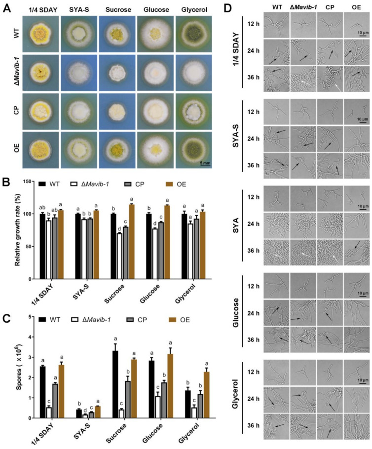
Conidium is the main infection unit and reproductive unit of pathogenic fungi. Exploring the mechanism of conidiation and its regulation contributes to understanding the pathogenicity of pathogenic fungi. Vib-1, a transcription factor, was reported to participate in the conidiation process. However, the regulation mechanism of Vib-1 in conidiation is still unclear. In this study, we analyzed the function of Vib-1 and its regulation mechanism in conidiation through knocking out and overexpression of Vib-1 in entomopathogenic fungus Metarhizium acridum. Results showed that the colonial growth of Mavib-1 disruption mutant (ΔMavib-1) was significantly decreased, and conidiation was earlier compared to wild type (WT), while overexpression of Mavib-1 led to a delayed conidiation especially when carbon or nitrogen sources were insufficient. Overexpression of Mavib-1 resulted in a conidiation pattern shift from microcycle conidiation to normal conidiation on nutrient-limited medium. These results indicated that Mavib-1 acted as a positive regulator in vegetative growth and a negative regulator in conidiation by affecting utilization of carbon and nitrogen sources in M. acridum. Transcription profile analysis demonstrated that many genes related to carbon and nitrogen source metabolisms were differentially expressed in ΔMavib-1 and OE strains compared to WT. Moreover, Mavib-1 affects the conidial germination, tolerance to UV-B and heat stresses, cell wall integrity, conidial surface morphology and conidial hydrophobicity in M. acridum. These findings unravel the regulatory mechanism of Mavib-1 in fungal growth and conidiation, and enrich the knowledge to conidiation pattern shift of filamentous fungi.

Keywords: carbon and nitrogen utilization; conidiation; entomopathogenic fungi; transcription factor Mavib-1.


Conflict of interest statement

The authors declare no conflict of interest.

Figures

Figure 1
Figure 1
Transcriptional-activation assays and subcellular localization of Mavib-1. (A) Transcriptional activation assays in yeast. Mavib-1-N (containing NDT80 domain), Mavib-1-C or Mavib-1 full CDS were fused to the GAL4 DNA-binding domain and expressed in yeast strain Y2H Gold. Strain containing only GAL4 DNA-binding domain was as negative control (NC), containing the GAL4 DNA-binding and activation domains were as positive controls (PC). All the yeast strains were cultured on SD-Trp plates, SD-Trp plates containing 0.5 mM X-α-gal and SD-Trp plates containing 0.5 mM X-α-gal and 200 ng/mL AbA (Aureobasidin A) at 30 °C for 3 days. (B) Subcellular localization of Mavib-1. The expression of Mavib-1 and EGFP fusion protein was driven by strong promoter PgpdM. The conidia and mycelia of OE strain were stained with DAPI (4′,6-diamidino-2-phenylindole, C0065, Solarbio, China), a fluorescent dye that can bind strongly to DNA, and observed under a confocal microscope (TCS SP8, Leica, Germany).
Figure 2
Figure 2
Stress tolerances of conidia from WT, ΔMavib-1, CP and OE strains. (A) Germination rate of each strain on 1/4 SDAY media at 28 °C for 20 h after UV-B stress treatment at 0, 1.25, 2.5, 3.75, 5.0 h. (B) The half inactivity time (IT50) of each strain with UV-B treatment. (C) Germination rates on 1/4 SDAY at 28 °C for 20 h after 46 °C heat stress treatment at 0, 3, 6, 9, 12 h. (D) The IT50 of WT, ΔMavib-1, CP and OE strains under 46 °C heat treatment. (E) The fungal colony on 1/4 SDAY medium and 1/4 SDAY with cell wall disruptors (500 µg/mL CR, 50 µg/mL CFW), cell wall stressor (0.01% SDS), hyperosmotic stressors (1 mol/L SOR or 1 mol/L NaCl), oxidative stress (6 mmol/L H2O2), respectively. (F) Inhibition rate of colony growth. All experiments were repeated three times for statistical analysis. Different capital letters represented significant difference at p < 0.01; different lower-case letters represented significant difference at p < 0.05.
Figure 3
Figure 3
Conidial germination, conidial surface morphology and hydrophobicity determination. (A) The 50% germination times (GT50) of WT, ΔMavib-1, CP and OE strains. (B) The conidial surface morphology by SEM. (C) Conidial hydrophobicity index. The trial was repeated three times. Different capital letters represented significant difference at p < 0.01 and different lower-case letters indicate significant difference, p < 0.05.
Figure 4
Figure 4
Colony growth and conidiation of fungal strains on media containing different carbon sources. (A) Colony morphology of WT, ΔMavib-1, CP and OE strains. Fungal strains were cultured on the nutrient-rich medium 1/4 SDAY, and nutrient-limited medium SYA with easy-to-use or difficult-to-use carbon sources (3% w/v; easy to use: glucose; middle: sucrose, difficult to use: glycerol) for 5 d. Colony relative growth rate (B) and conidial yield (C) of WT, ΔMavib-1, CP and OE strains cultured on 1/4 SDAY or SYA with different carbon sources for 5 d. The trial was repeated three times. Error bars are standard deviations of three replicates. Different lower-case letters indicate significant difference at p < 0.05. (D) Conidiation of WT, ΔMavib-1, CP and OE strains on different media. The black arrows indicate normal conidiation, and the white arrows indicate microcycle conidiation.
Figure 5
Figure 5
Colony growth and conidiation of ΔMavib-1, CP and OE strains on media with different nitrogen sources. Colony morphology (A) and colony relative growth rate (B) of ΔMavib-1, CP and OE strains on the media 1/4 SDAY, and SYA with different nitrogen sources. (C) Conidial yield of fungal strains cultured on different media for 15 d. Media contained 0.3% nitrogen source: SYA-N (SYA without NaNO3), urea (difficult to use), NaNO3 (middle), Gln (easy to use). Each trial was repeated three times for statistical analysis. Different lower-case letters mean significant difference at p < 0.05. (D) Conidiation of ΔMavib-1, CP and OE strains on different media. The black arrows indicate NC, and the white arrows indicate MC.
Figure 6
Figure 6
DEGs analysis. (A) Statistical histogram of the number of DEGs. (B) Venn diagram of DEGs. (C) Heat map of 18 DEGs downregulated in ΔMavib-1 and upregulated in OE. The 18 DEGs consistent with the change trend of Mavib-1 were analyzed by cluster analysis of expression patterns. The distance calculation method was used: the spearman correlation coefficient between samples, the Pearson correlation coefficient between genes. (D) Cluster heat map of the expression levels of the 4 genes upregulated in ΔMavib-1 and downregulated in OE. (E) Heat map of the expression of DEGs in ΔMavib-1 annotated as CAZyme genes. (F) Heat map of the expression of DEGs in OE annotated as CAZyme genes. The up and down arrows represent upregulated or downregulated genes, respectively. Numbers in blue represent ΔMavib-1 and red represent OE. The red color denotes the upregulated DEGs, and the blue color denotes downregulated DEGs. The log2 (expression value + 1) of the sample were shown in the horizontal axis, and DEGs are shown at the right-hand vertical side.

Similar articles

Cited by

References

    1. Pendland J.C., Hung S.Y., Boucias D.G. Evasion of host defense by in vivo-produced protoplast-like cells of the insect mycopathogen Beauveria bassiana. J. Bacteriol. 1993;175:5962–5969. doi: 10.1128/jb.175.18.5962-5969.1993. - DOI - PMC - PubMed
    1. Khurana N., Saxena R.K., Gupta R., Kuhad R.C. Light-independent conidiation in Trichoderma spp.: A novel approach to microcycle conidiation. World J. Microbiol. Biotechnol. 1993;9:353–356. doi: 10.1007/BF00383079. - DOI - PubMed
    1. Clarkson J.M., Charnley A.K. New insights into the mechanisms of fungal pathogenesis in insects. Trends Microbiol. 1996;4:197–203. doi: 10.1016/0966-842X(96)10022-6. - DOI - PubMed
    1. Zhang S.Z., Peng G.X., Xia Y.X. Microcycle conidiation and the conidial properties in the entomopathogenic fungus Metarhizium acridum on agar medium. Biocontrol Sci. Technol. 2010;20:809–819. doi: 10.1080/09583157.2010.482201. - DOI
    1. De Faria M.R., Wraight S.P. Mycoinsecticides and mycoacaricides: A comprehensive list with worldwide coverage and international classification of formulation types. Biol. Control. 2007;43:237–256. doi: 10.1016/j.biocontrol.2007.08.001. - DOI

LinkOut - more resources