Review
. 2022 Jun 10;8(6):621.
doi: 10.3390/jof8060621.

Post-Translational Modifications of PCNA: Guiding for the Best DNA Damage Tolerance Choice

Affiliations
Review

Post-Translational Modifications of PCNA: Guiding for the Best DNA Damage Tolerance Choice

Gemma Bellí et al. J Fungi (Basel). .

Abstract

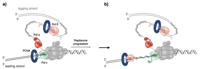
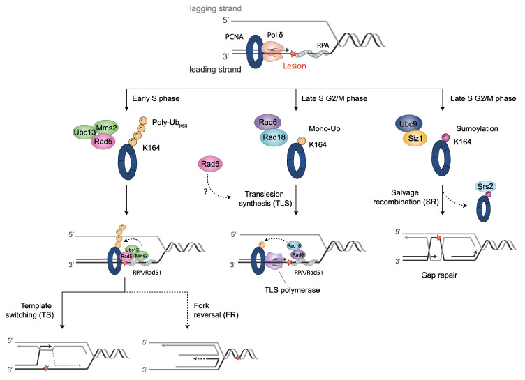
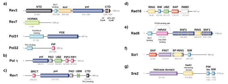
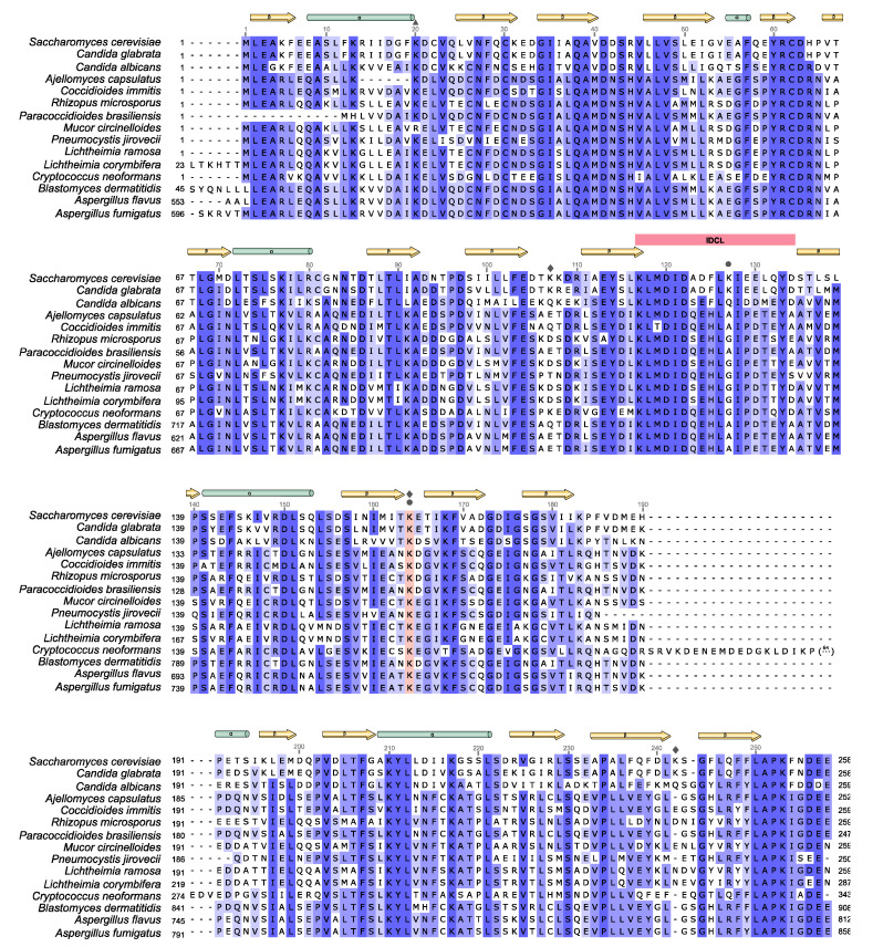
The sliding clamp PCNA is a multifunctional homotrimer mainly linked to DNA replication. During this process, cells must ensure an accurate and complete genome replication when constantly challenged by the presence of DNA lesions. Post-translational modifications of PCNA play a crucial role in channeling DNA damage tolerance (DDT) and repair mechanisms to bypass unrepaired lesions and promote optimal fork replication restart. PCNA ubiquitination processes trigger the following two main DDT sub-pathways: Rad6/Rad18-dependent PCNA monoubiquitination and Ubc13-Mms2/Rad5-mediated PCNA polyubiquitination, promoting error-prone translation synthesis (TLS) or error-free template switch (TS) pathways, respectively. However, the fork protection mechanism leading to TS during fork reversal is still poorly understood. In contrast, PCNA sumoylation impedes the homologous recombination (HR)-mediated salvage recombination (SR) repair pathway. Focusing on Saccharomyces cerevisiae budding yeast, we summarized PCNA related-DDT and repair mechanisms that coordinately sustain genome stability and cell survival. In addition, we compared PCNA sequences from various fungal pathogens, considering recent advances in structural features. Importantly, the identification of PCNA epitopes may lead to potential fungal targets for antifungal drug development.

Keywords: DNA damage tolerance; DNA replication forks; DNA replication stress; PCNA; fungal genome stability; post-translational modifications; salvage recombination; template switch; translesion synthesis.


Conflict of interest statement

The authors declare no conflict of interest.

Figures

Figure 1
Figure 1
The polymerase switch facilitates the bypass of DNA lesions on the leading strand. (a) During unperturbed replisome progression, leading and lagging strands are synthesized by Polε and Polα/Polδ, respectively. (b) When the replisome encounters a DNA lesion that Polε is not able to tolerate, Polδ plays a key role in the initiation of leading-strand synthesis. The Cdc45-Mcm2-7-GINS (CMG) helicase complex is depicted in grey. Blocking DNA lesion is depicted as a red star.
Figure 2
Figure 2
PCNA post-translational modifications regulate DDT pathways. When fork stalling persists, cells activate DDT mechanisms through post-translational modifications on PCNA. Monoubiquitination of PCNA at K164 by Rad6-Rad18 promotes the switch from Polδ to translesion synthesis (TLS) polymerases for error-prone TLS damage bypass. K63 extended polyubiquitination of PCNA on K164 by Mms2/Ubc13-Rad5 for error-free damage bypass mediates template switching (TS). This modification might be also implied in fork protection by fork reversal. Unloading of SUMO-PCNA bound to Srs2 (sumoylated by Ubc9-Siz1) provides the salvage recombination pathway (SR) alternatively to TS or TLS, either at stalled replication forks (RFs) or, as shown, at gaps left behind RFs after re-initiation. Cell cycle stages where DDT processes predominantly take place in yeast are indicated. Blocking DNA lesions are depicted as a red star.
Figure 3
Figure 3
Structure organization of PCNA-interacting mediators involved in DDT in S. cerevisiae: TLS polymerases (ac), modifying-E3 enzymes (df) and Srs2 helicase (e). (a) Subunits of Polζ. The catalytic subunit, Rev3, contains an inactive 3’-5’ exonuclease domain (exo-), a DNA polymerase domain (pol), and a conserved CysA and CysB sites in its C-terminal domain (CTD), containing a Zinc Finger (ZF) and a [4Fe-4S] cluster, respectively. The Rev7 binding site is located towards its N-terminal domain (NTD). Rev7 contains the Hop1, Rev7 and Mad2 family domain (HORMA). Pol31 contains a Rev3/Pol3 binding site and a phosphodiesterase domain (PDE). Pol32 binds Pol1 and contains a PCNA-interactive motif (PIP). (b) Polη includes pol and PAD domains in its NTD, a ubiquitin-binding zinc finger motif (UBZ), and the PIP1 and PIP2 motifs at the CTD. (c) Rev1 contains a pol domain, a polymerase associated domain (PAD), two small ubiquitin binding motifs (UBM), a small rev7 binding domain CTD and a BRCA1 NTD (BRCT). (d) E3 Ub-ligase Rad18 contains a RING (Really Interesting New Gene) domain, the SUMO interacting motif (SIM), the UBZ motif, SAF-A/B, Acinus, Pias (SAP) domain, and Rad6-Binding Domain (R6BD). (e) E3 Ub-ligase Rad5 contains the Rev1 binding domain, a HIRAN domain (HIP116 Rad5p N-terminal), the helicase domain (SNF2), and a RING domain. (f) E3 SUMO-ligase Siz1 includes SAP, PINT and SP-RING domains, and the SIM motif. (g) Srs2 helicase contains the helicase domain at its NTD, a Rad51 interacting domain, and a PIM and a SIM motif at CTD. The name and length (number of amino acids) of each PCNA-binding protein are indicated.
Figure 4
Figure 4
Alignment of multiple PCNA sequences of fungal pathogens, causing systemic infections, compared to S. cerevisiae. Multiple sequences alignment of S. cerevisiae PCNA and PCNA from 14 pathogenic fungal species is shown. S. cerevisiae (P15873), Candida glabrata (Q6FWA4), Candida albicans (Q5AMN0), Ajellomyces capsulatus (A6R5C7), Coccidioides immitis (A0A0J7B5C4), Rhizopus microsporus (A0A0A1P4Z3), Paracoccidioides brasiliensis (A0A1D2J4G1), Mucor circinelloides (S2K5N0), Pneumocystis jirovecii (A0A0W4ZHN6), Lichtheimia ramosa (A0A077WRZ8), Lichtheimia corymbifera (A0A068SE94), Cryptococcus neoformans (Q5K7Y2), Blastomyces dermatitidis (T5B6A2), Aspergillus flavus (B8N1A6), Aspergillus fumigatus (A0A0J5SJF1). PCNA sequences were obtained using Uniprot repository database (Uniprot entries are indicated in parentheses), and the sequence alignment was carried out using UGene software. Identical residues are shaded dark blue, whereas similar residues are shaded light blue. Secondary structural features are indicated above the sequences alignment, α-helices (yellow) and β-strands (green). Conserved IDCL motifs and K164 residues are shaded pink. The symbol on the upper part of the alignment indicates lysine modification—triangle for acetylation; rhombus for ubiquitylation; and circle for SUMOylation.

Similar articles

Cited by

References

    1. Bauer G.A., Burgers M.J. Molecular cloning, structure and expression of the yeast proliferating cell nuclear antigen gene. Nucleic Acids Res. 1990;18:261–265. doi: 10.1093/nar/18.2.261. - DOI - PMC - PubMed
    1. Waga S., Stillman B. The DNA replication fork in eukaryotic cells. Annu. Rev. Biochem. 1998;67:721–751. doi: 10.1146/annurev.biochem.67.1.721. - DOI - PubMed
    1. Moldovan G.L., Pfander B., Jentsch S. PCNA, the Maestro of the Replication Fork. Cell. 2007;129:665–679. doi: 10.1016/j.cell.2007.05.003. - DOI - PubMed
    1. Henderson D.S., Banga S.S., Grigliatti T.A., Boyd J.B. Mutagen sensitivity and suppression of position-effect variegation result from mutations in mus209, the Drosophila gene encoding PCNA. EMBO J. 1994;13:1450–1459. doi: 10.1002/j.1460-2075.1994.tb06399.x. - DOI - PMC - PubMed
    1. Nomura A. Nuclear distribution of proliferating cell nuclear antigen (PCNA) in fertilized eggs of the starfish Asterina pectinifera. J. Cell Sci. 1994;107:3291–3300. doi: 10.1242/jcs.107.12.3291. - DOI - PubMed

LinkOut - more resources