. 2022 Jul 8;50(12):6656-6670.
doi: 10.1093/nar/gkac539.

From bench to bedside: Improving the clinical safety of GalNAc-siRNA conjugates using seed-pairing destabilization

Affiliations

From bench to bedside: Improving the clinical safety of GalNAc-siRNA conjugates using seed-pairing destabilization

Mark K Schlegel et al. Nucleic Acids Res. .

Abstract

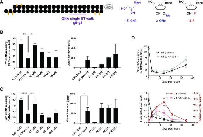
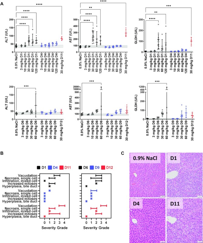
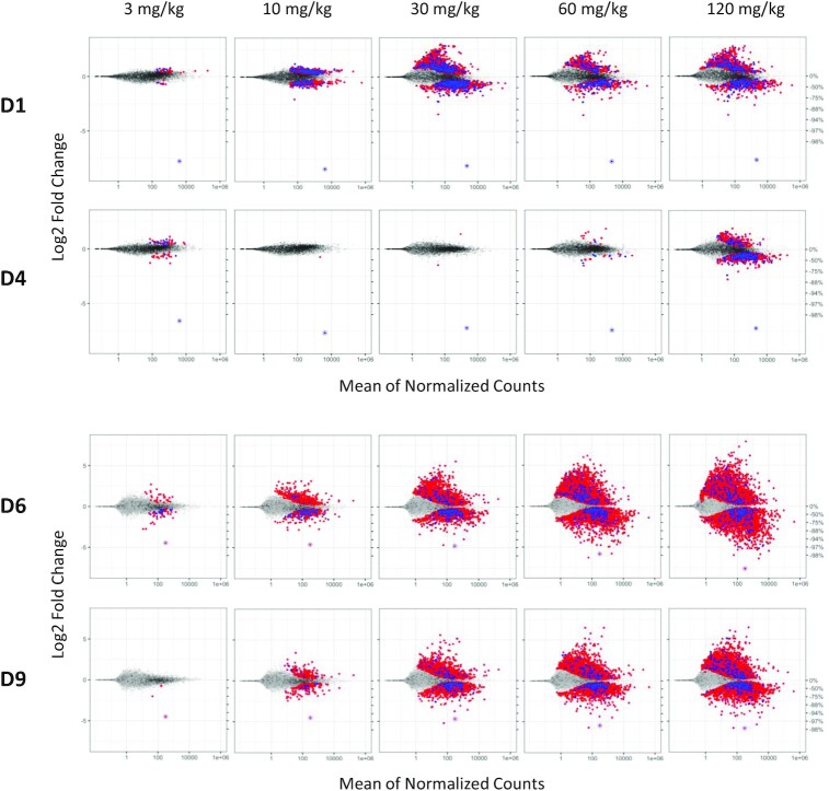
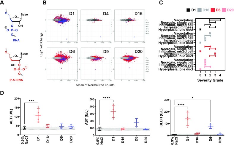
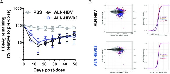
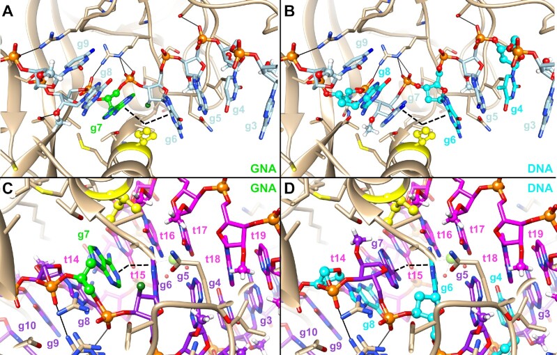
Preclinical mechanistic studies have pointed towards RNA interference-mediated off-target effects as a major driver of hepatotoxicity for GalNAc-siRNA conjugates. Here, we demonstrate that a single glycol nucleic acid or 2'-5'-RNA modification can substantially reduce small interfering RNA (siRNA) seed-mediated binding to off-target transcripts while maintaining on-target activity. In siRNAs with established hepatotoxicity driven by off-target effects, these novel designs with seed-pairing destabilization, termed enhanced stabilization chemistry plus (ESC+), demonstrated a substantially improved therapeutic window in rats. In contrast, siRNAs thermally destabilized to a similar extent by the incorporation of multiple DNA nucleotides in the seed region showed little to no improvement in rat safety suggesting that factors in addition to global thermodynamics play a role in off-target mitigation. We utilized the ESC+ strategy to improve the safety of ALN-HBV, which exhibited dose-dependent, transient and asymptomatic alanine aminotransferase elevations in healthy volunteers. The redesigned ALN-HBV02 (VIR-2218) showed improved specificity with comparable on-target activity and the program was reintroduced into clinical development.


Figures

Figure 1.
Figure 1.
Evaluation of GNA-modified siRNAs in mice. (A) Structures of (S)-GNA, 2′-OMe and 2′-F nucleosides. Impact of GNA substitution at the specified position on activity (left) and liver guide strand concentration measured by RT-qPCR (right) in mice (n = 3) 7 days post-dose for Ttr-targeting (B) or Hao1-targeting (C) GalNAc–siRNAs dosed subcutaneously at either 0.5 mg/kg (Ttr) or 1.0 mg/kg (Hao1). Statistically significant differences relative to the parent siRNA are shown in each graph. (D) Impact of GNA substitution on Ttr mRNA knockdown in mice (n = 3) after a single subcutaneous dose of 0.75 mg/kg D1 or D4 and guide strand concentration in whole liver and Ago2 over time from the same study.
Figure 2.
Figure 2.
Selected clinical pathology parameters and microscopic liver findings measured in rats (n = 4–5) following three weekly doses of parent, GNA- or DNA-modified GalNAc–siRNAs targeting Ttr or Hao1. (A) Measured liver enzymes from serum that was collected 24 h after final dose. Data were collected from three different studies; D1, D4, D6 and D9 were evaluated in a single study at 3, 10 and 30 mg/kg, and two subsequent studies evaluated D1 and D4 or D6 and D9 at 30, 60 and 120 mg/kg, and D11 or D12 at 30 mg/kg. Controls and the overlapping 30 mg/kg groups were combined and plotted above. (B) Summary of the range of microscopic liver findings based on severity grade. (C) Microscopic liver findings in rats following three weekly doses of 30 mg/kg with the indicated siRNAs targeting Ttr compared to the 0.9% NaCl control. Livers were collected 24 h post-final dose for analysis and sections were stained with H&E.
Figure 3.
Figure 3.
Transcriptional dysregulation in rat livers following three weekly doses of GalNAc–siRNAs targeting Ttr (D1, D4, top) or Hao1 (D6, D9, bottom). Frozen livers were collected 24 h after last dose for RNA-seq analysis. Log2 fold change plots (MA plot) represent the average signal from each cohort (n = 4–5). Dots represent individual rat gene transcripts, their average read count and the level of change in expression compared to the control group dosed with 0.9% NaCl. Whereas gray dots represent gene transcripts that were not determined to be differentially expressed after siRNA treatment relative to the control, the blue and red dots represent differentially expressed gene transcripts (false discovery rate < 0.05) with or without a canonical miRNA match (8mer, 7mer-m8, 7mer-A1 and mer6) to the guide seed region, respectively. On-target knockdown is represented by the circled dot.
Figure 4.
Figure 4.
Evaluation of 2′–5′-RNA-modified siRNAs in rats. (A) Structures of RNA and 2′–5′-RNA nucleotides. (B) Transcriptional dysregulation in primary rat hepatocytes following transfection of the specified siRNAs at a dose of 50 nM (n = 4). (C) Summary of the range of microscopic liver findings based on severity grade after three weekly doses of 30 mg/kg in rats (n = 4). (D) Measured liver enzymes from serum that was collected 24 h after final dose.
Figure 5.
Figure 5.
Evaluation of the efficacy and specificity of ALN-HBV02. (A) Pharmacodynamics after a single 1 mg/kg dose of ALN-HBV or ALN-HBV02 in mice transduced with HBV-AAV8. Serum HBsAg levels represent the average and error bars represent the standard deviation of all animals from a given cohort (n = 3), each normalized to individual pre-dose serum HBsAg levels. (B) Measurement of transcriptional dysregulation in HepG2.2.15 after transfection at 10 nM with ALN-HBV or ALN-HBV02 relative to a mock control. MA and CDF plots are shown on the left and right, respectively. In CDF plots, each colored line represents the impact of different types of seed matches on the cumulative dysregulation of gene transcripts: black = background; purple = mer6; yellow = mer7-A1; blue = mer7-m8; and red = mer8.
Figure 6.
Figure 6.
Modeling of modified guide strands in human Ago2. GNA-modified guide strand of D4 (A) or DNA-modified guide strand of D11 (B) modeled into the structure of Ago2–guide complex (PDB code 4F3T) (41). The angle of the kink in the guide strand introduced by Ile365 is indicated by the black dashed wedge. The same guide strands, now modeled into a structure of Ago2 in complex with both guide and target RNAs (PDB code 4W5T), are shown in panels (C) and (D) (7). A relax of the kink between g6 and g7 can be observed with a close to A-like conformation of the guide–target duplex.

References

    1. Nair J.K., Willoughby J.L.S., Chan A., Charisse K., Alam M.R., Wang Q., Hoekstra M., Kandasamy P., Kel’in A.V., Milstein S.et al. .. Multivalent N-acetylgalactosamine-conjugated siRNA localizes in hepatocytes and elicits robust RNAi-mediated gene silencing. J. Am. Chem. Soc. 2014; 136:16958–16961. - PubMed
    1. Prakash T.P., Graham M.J., Yu J., Carty R., Low A., Chappell A., Schmidt K., Zhao C., Aghajan M., Murray H.F.et al. .. Targeted delivery of antisense oligonucleotides to hepatocytes using triantennary N-acetyl galactosamine improves potency 10-fold in mice. Nucleic Acids Res. 2014; 42:8796–8807. - PMC - PubMed
    1. Zimmermann T.S., Karsten V., Chan A., Chiesa J., Boyce M., Bettencourt B.R., Hutabarat R., Nochur S., Vaishnaw A., Gollob J.. Clinical proof of concept for a novel hepatocyte-targeting GalNAc–siRNA conjugate. Mol. Ther. 2017; 25:71–78. - PMC - PubMed
    1. Foster D.J., Brown C.R., Shaikh S., Trapp C., Schlegel M.K., Qian K., Sehgal A., Rajeev K.G., Jadhav V., Manoharan M.et al. .. Advanced siRNA designs further improve in vivo performance of GalNAc–siRNA conjugates. Mol. Ther. 2018; 26:708–717. - PMC - PubMed
    1. Nair J.K., Attarwala H., Sehgal A., Wang Q., Aluri K., Zhang X., Gao M., Liu J., Indrakanti R., Schofield S.et al. .. Impact of enhanced metabolic stability on pharmacokinetics and pharmacodynamics of GalNAc–siRNA conjugates. Nucleic Acids Res. 2017; 45:10969–10977. - PMC - PubMed

Publication types

Substances

LinkOut - more resources