. 2022 Jul 8;50(12):7134-7146.
doi: 10.1093/nar/gkac523.

G-quadruplex structure of the C. elegans telomeric repeat: a two tetrads basket type conformation stabilized by a non-canonical C-T base-pair

Affiliations

G-quadruplex structure of the C. elegans telomeric repeat: a two tetrads basket type conformation stabilized by a non-canonical C-T base-pair

Julien Marquevielle et al. Nucleic Acids Res. .

Abstract

The Caenorhabditis elegans model has greatly contributed to the understanding of the role of G-quadruplexes in genomic instability. The GGCTTA repeats of the C. elegans telomeres resemble the GGGTTA repeats of the human telomeres. However, the comparison of telomeric sequences (Homo sapiens, Tetrahymena, Oxytricha, Bombyx mori and Giardia) revealed that small changes in these repeats can drastically change the topology of the folded G-quadruplex. In the present work we determined the structure adopted by the C. elegans telomeric sequence d[GG(CTTAGG)3]. The investigated C. elegans telomeric sequence is shown to fold into an intramolecular two G-tetrads basket type G-quadruplex structure that includes a C-T base pair in the diagonal loop. This work sheds light on the telomeric structure of the widely used C. elegans animal model.


Figures

Graphical Abstract
Graphical Abstract
The GGCTTA repeats of the C. elegans telomeres, similar to the GGGTTA repeats in human telomeres, fold into an intramolecular two G-tetrads basket type G-quadruplex structure including a C–T base pair.
Figure 1.
Figure 1.
Reported G4 topologies for telomeric sequences from (A–H) human (GGGTTA repeat or GGGCTA repeat for (H)) and non-human (I–L) organisms. Syn and anti guanines are represented in magenta and cyan, respectively, while cytosines are shown in green. W, N and M refer to wide, narrow and medium grooves, respectively. Each topology is described using the single descriptor nomenclature (SDN) (51) containing the number of guanines in the stem (n = 2, 3 or 4) along with the type of loops (lw: lateral spanning a wide groove, ln: Lateral spanning a narrow groove, p: propeller and d for diagonal) and relative direction (+ or –) of loops linking G-tracts of the stem: n(L1, L2,L3). (A) 2+2 conformation in Na+ (pdb: 2MBJ) (19,20). SDN: 3(+Ln-P-Lw). (B) 2+2 basket type conformation in Na+ (pdb: 143D) (19,20). SDN: 3(−LwD+Ln). (C) 3+1 conformation in K+ (pdf: 2JPZ) (21–26). SDN: 3(−Lw−Ln−P). (D) 3+1 conformation in K+ (pdb: 2GKU) (21–26). SDN: 3(−P−Ln−Lw). (E) All parallel conformation (pdb: 2LD8) (21–26) SDN: 3(-P-P-P). (F) 2+2 basket type conformation in K+ (pdb: 2KF8 & 2KKA (55)) (21–26). SDN: 2(−LwD+Ln). (G) 2+2 basket type conformation in K+ (pdb: 5LQG) with only 3 telomeric repeats (27). SDN: 2(+LnD−Lw). (H) Chair type Anti-parallel conformation for the human variant in K+ (pdb: 2KM3) (28). SDN: 2(+Ln+Lw+Ln). (I) 2+2 basket type conformation for Oxytricha (G4T4)n in Na+ (pdb: 201D) (13). SDN: 4(−LwD+Ln). (J) Chair type Anti-parallel conformation for Bombyx mori (G2TTA)n in K+ (15). SDN: 2(-Lw-Ln-Lw). (K) 2+2 basket type conformation for Giardia (G3TA)n in K+ (pdb: 2KOW) (16). SDN: 2(+LnD−Lw). (L) 3+1 conformation for Tetrahymena (G4T2)n in Na+ (pdb: 186D) (13). SDN: 3(−Lw−Ln−P).
Figure 2.
Figure 2.
(A–C) Optical spectroscopy on a 5 μM Ce20 sample in 20 mM potassium phosphate buffer pH 6.9 supplemented with 70 mM KCl. (A) Thermal difference spectrum (TDS) (B) UV-melting (both heating and cooling profiles are shown; Tm = 40°C) (C) CD-spectrum. (D) 1H NMR spectrum at 288 K in 90% H2O/10% D2O 20 mM potassium phosphate buffer pH 6.9 supplemented with 70 mM KCl. (*) Guanine imino protons from tetrads, (+) Thymin imino proton, (Δ) guanine amino proton.
Figure 3.
Figure 3.
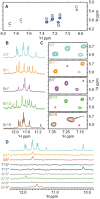
Unambiguous assignment of Imino/aromatic protons of guanines based on 15N-labelling and Br-guanine substitutions. Experiments were recorded in 90% H2O /10% D2O 20 mM potassium phosphate buffer pH 6.9 supplemented with 70 mM KCl. (A) {1H–1H} NOESY spectrum of Ce20 with a 50 ms mixing time. (B, C) 1H 1D NMR spectra of Ce20, Br1, Br7, Br13 and Br19 at 288 K with in (B) 1D spectra and in (C) NOESY experiments with a mixing time of 50 ms). (D) 15N-filtrated spectra using samples containing 5% of 15N-enriched isotope in order determine or confirm residues implicated in Ce20 G4 formation.
Figure 4.
Figure 4.
Assignment of aromatic and imino protons of guanines in 90% H2O /10% D2O 20 mM potassium phosphate buffer pH 6.9 supplemented with 70 mM KCl. (A) {13C−1H}-HMBC at 302 K showing intra-residue H1/H8 correlations used to identify H8 of guanines implicated in Ce20 tetrads. (B) Ce20 inter-residue H1’/H8 correlations (“walk”) between H8 from a residue and H1’ from the previous residue {1H–1H} NOESY experiment with a 250 ms mixing time. Red stars represent weak correlations.
Figure 5.
Figure 5.
Determination of the G-quadruplex topology in 90% H2O /10% D2O 20 mM potassium phosphate buffer pH 6.9 supplemented with 70 mM KCl. (A) and (B) Inter-residue H1/H1 and H1/H8 correlations used to determine tetrads patterns in {1H–1H} NOESY experiment with a 250 ms mixing time. Tetrads patterns and Ce20 topology are respectively shown in (C) and (D). Syn and anti guanines are represented in magenta and cyan, respectively, while cytosines are shown in green. W, N and M refer to wide, narrow and medium grooves, respectively.
Figure 6.
Figure 6.
Structure of the Ce20 G-quadruplex. (A) Ensemble of the ten best structures obtained after structure calculation and refinement. Guanines are coloured in green, Adenines in cyan, cytosines in purple and thymines in orange (B) Surface view of the ensemble. Specific features of Ce20 G-quadruplex structure are shown in (C) with the stacking of A18 (cyan) on top of G18 and the C9–T11 base pair (purple and orange) at the bottom of the bottom tetrad. (D) Top view of the C9–T11 base pair that stacks on top of G1 and G8. (E) Top view of the A18 that stacks on top of the G2–G7–G19–G14 tetrad.
Figure 7.
Figure 7.
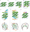
Topologies of 2-quartet intramolecular G-quadruplexes. Guanine tetrads are represented in dark green, while guanines involved in base-pairs or triads are shown in light green. Cytosines, Thymines and Adenines are represented in violet, yellow and blue. W, N and M refer to wide, narrow and medium grooves, respectively. Each topology is described using the single descriptor nomenclature (51) containing the number of guanines in the stem (n = 2) along with the type of loops (lw: Lateral spanning a wide groove, ln: Lateral spanning a narrow groove and d for diagonal) and relative direction (+ or –) of loops linking G-tracts of the stem: n(L1, L2,L3). (A–D) Chair type anti-parallel conformation for: (A) Bombyx mori (G2TTA)n in K+ (15). SDN: 2(-Lw-Ln-Lw). (B) The thrombin binding aptamer d[GGTTGGTGTGGTTGG] (PDB: 148D). SDN: 2(+Ln+Lw+Ln). (C) The HIV-PRO1 sequence d[TGGCCTGGGCGGGACTGGG] (56). SDN: 2(-Lw-Ln-Lw). (D) The telomeric human variant in K+ containing a G:C:G:C tetrad (pdb: 2KM3) (28). SDN: 2(+Ln+Lw+Ln). (E, F) Basket type anti-parallel conformation for: (E) The telomeric motif of Giardia (G3TA)n in K+ (pdb: 2KOW) (16). SDN: 2(+LnD−Lw). (F) The telomeric motif of Human in K+ (pdb = 2KF8 & 2KKA(55)) (21–26). SDN: 2(−LwD+Ln). (G) The telomeric motif of C. elegans (this work; PDB: 7OQT). SDN: 2(−LwD+Ln).

References

    1. Teixeira M.T. Saccharomyces cerevisiae as a model to study replicative senescence triggered by telomere shortening. Front. Oncol. 2013; 3:101–101. - PMC - PubMed
    1. Tran P.L.T., Mergny J.-L., Alberti P.. Stability of telomeric G-quadruplexes. Nucleic Acids Res. 2011; 39:3282–3294. - PMC - PubMed
    1. Maizels N. G4-associated human diseases. EMBO Rep. 2015; 16:910–922. - PMC - PubMed
    1. Bryan T.M. G-Quadruplexes at telomeres: friend or foe. Mol. 2020; 25:3686. - PMC - PubMed
    1. Postberg J., Tsytlonok M., Sparvoli D., Rhodes D., Lipps H.J.. A telomerase-associated RecQ protein-like helicase resolves telomeric G-quadruplex structures during replication. Gene. 2012; 497:147–154. - PMC - PubMed

Publication types