Review
. 2022 Jul 8;50(12):6618-6638.
doi: 10.1093/nar/gkac519.

Anatomy of four human Argonaute proteins

Affiliations
Review

Anatomy of four human Argonaute proteins

Kotaro Nakanishi. Nucleic Acids Res. .

Abstract

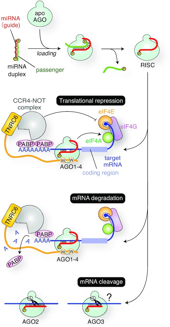
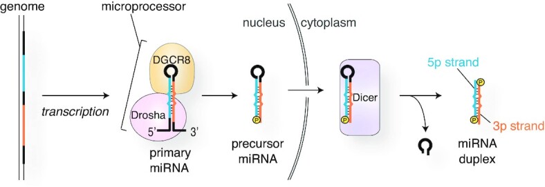
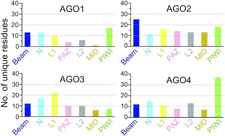
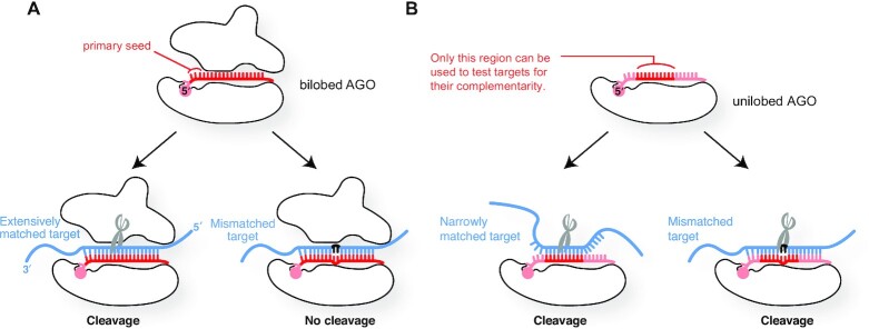
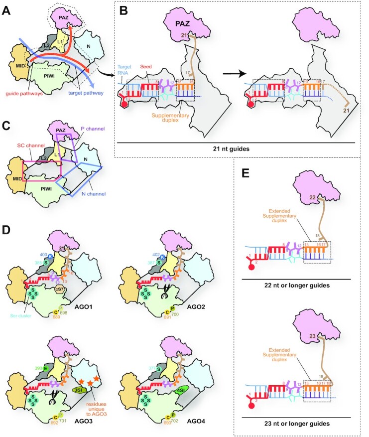
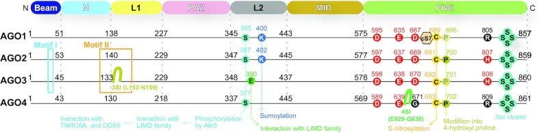
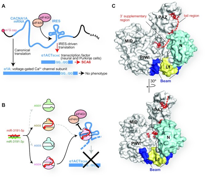
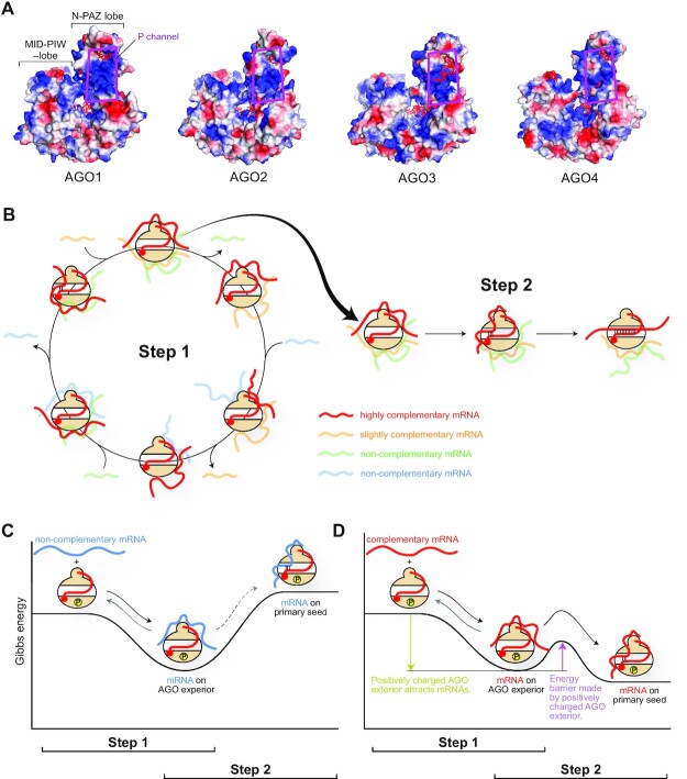
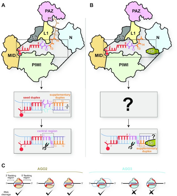
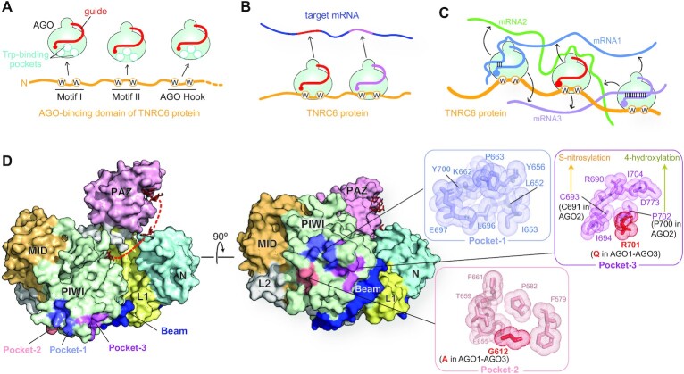
MicroRNAs (miRNAs) bind to complementary target RNAs and regulate their gene expression post-transcriptionally. These non-coding regulatory RNAs become functional after loading into Argonaute (AGO) proteins to form the effector complexes. Humans have four AGO proteins, AGO1, AGO2, AGO3 and AGO4, which share a high sequence identity. Since most miRNAs are found across the four AGOs, it has been thought that they work redundantly, and AGO2 has been heavily studied as the exemplified human paralog. Nevertheless, an increasing number of studies have found that the other paralogs play unique roles in various biological processes and diseases. In the last decade, the structural study of the four AGOs has provided the field with solid structural bases. This review exploits the completed structural catalog to describe common features and differences in target specificity across the four AGOs.


Figures

Figure 1.
Figure 1.
RISC assembly and the gene silencing pathways. AGO incorporates a miRNA duplex, ejects the passenger strand (green), and forms the RISC with the remaining guide strand (red). The effector complexes of all four human AGOs cause translational repression and mRNA degradation. Meanwhile, only AGO2 and AGO3 become a slicer, though target RNAs cleaved by AGO3-RISC remain unknown.
Figure 2.
Figure 2.
Numbering system of nucleotides on guide and target strands. (A) A guide RNA can be split into four regions, seed (red), central (magenta), 3′ supplementary (orange) and tail (wheat). (B) Nucleotides on target RNAs (bottom strand) are numbered based on the paired nucleotide on the guide RNA (top strand).
Figure 3.
Figure 3.
Update on the AGO structure. (A) Domain architecture of human AGO. The residue numbers are those of AGO2. Six Loops (dark green) in the two PlWI subdomains are shown on the lower right side. (B) Beam running along the bottom of AGO. The color code is the same in Panel A. (C) PIWI-helical subdomain (ribbon model) mediates the MID domain (wheat) and PIWI-catalytic subdomain (white) in human AGO4 (left, PDB ID: 6OON). The disorder of a PIWI-helical subdomain of T. thermophilus AGO is highlighted with the red dotted circle (right, PDB ID: 3DLB). (D) Recognition of the 5′ monophosphate group by the AGO4 MID domain and PIWI-helical subdomain. All residues shown as stick models here are conserved across the four human AGOs. Water molecules are depicted as blue spheres. (E) Three Loops of the PIWI domain forming the seed-binding site. The C-terminal serine cluster is located on Loop 5.
Figure 4.
Figure 4.
Water molecules underpinning the seed-binding site. (A) Surface model (left) and cross-section (right) of human AGO4 in complex with guide RNA (red) (PDB ID: 6OON). Water molecules (blue spheres) forming LAKE1 and LAKE2 are buried inside the AGO4 (right). (B) The loops forming the seed-binding site are glued together by LAKE1 and LAKE2. The domain color codes are the same in Figure 3B. The trapped water molecules are depicted as cyan spheres. (C) Model of the water-mediated RISC assembly.
Figure 5.
Figure 5.
Asymmetric guide selection. (A) A strand with a thermodynamically unstable 5′ end (red strand) is incorporated as the guide into AGO. The thermodynamically stable and unstable ends are highlighted with cyan and orange blurs, respectively. The MID and PAZ domains are colored in wheat and pink, respectively. A 5′ monophosphate group is shown as ‘P’ in a circle. (B, C) Schematics of the crystal structures of PAZ-duplex complex (B) (23) and T. thermophilus AGO in complex with guide RNA (C) (29). (D) The currently accepted mechanism of asymmetric guide selection. The overall structure of AGO during RISC assembly remains unclear. (E) The PAZ domain binds to either of the duplex termini, resulting in the binding of the MID domain to the other end. In the bottom pathway, when the PAZ domain binds to the thermodynamically unstable end (the 3′ end of the green strand), it remains unclear how the MID domain can recognize the 5′ end of the red strand and take the strand as a guide.
Figure 6.
Figure 6.
miRNA processing pathway.
Figure 7.
Figure 7.
The number of residues unique to each AGO per domain.
Figure 8.
Figure 8.
Roles of the N-PAZ lobe. (A) The bilobed structure is essential for high target specificity and mismatch recognition. (B) The lack of the N-PAZ lobe impairs the target specificity and mismatch recognition.
Figure 9.
Figure 9.
Differences in the N channel between four human AGOs. (A) Pathways of guide and target strands. The domain color codes are the same as in Figure 3. The nucleic acid-binding cleft is colored in gray. (B) Transfer of the guide 3′ supplementary and tail regions between two branched channels. In the case of a 21 nt guide, the g2–g8 and g13–g16 serve as the target binding sites. (C) Y-shaped nucleic acid-binding cleft. The cleft is composed of the Seed-Central channel (SC channel in the red box), the PAZ channel (P channel in the magenta box), and the N channel (blue box). (D) Local structures unique to each AGO. cS7, 3SI and 4SI are localized in the N channel. The catalytic DEDH tetrad is depicted as scissors. AGO3-specific residues on its N domain are shown as orange stars. The residues undergoing post-translational modification are shown with their residue number (see Figure 10). (E) Model of expanding the 3′ supplementary region. When the guide lengths are 22 nt (top) and 23 nt (bottom), the g17 and the g17–g18 participate in target recognition as part of the 3′ supplementary region, respectively.
Figure 10.
Figure 10.
Locations of the motifs, insertions, and modification sites. Red circles are the catalytic tetrad. Numbers in black indicate the first residue on each domain.
Figure 11.
Figure 11.
Unique roles of AGO4. (A) IRES-driven translation of CACNA1A mRNA causes spinocerebellar ataxia 6 when the mRNA includes polyglutamine repeats. (B) Both AGO2 and AGO4 load miR-3191–5p, but only the AGO4-RISC stops the IRES-driven translation. (C) The N-terminal regions of AGO4 highlighted in cyan, yellow and blue serve as the DNMT3A-binding site. The bound guide RNAs are depicted as red stick models (PDB ID: 6OON).
Figure 12.
Figure 12.
Bifunctional positively charge exterior of AGO. (A) Electrostatic potential map of four human AGOs. Positive and negative potentials are drawn in blue and red, respectively. The P channel is highlighted with magenta boxes. (B) Model of two-step RNA recognition by AGOs. Slightly- and non-complementary mRNAs are made fainter for clarity. (C, D) Energy levels of non-complementary mRNA (C) and complementary mRNA (D). AGO is phosphorylated to some extent.
Figure 13.
Figure 13.
Differences in target recognition between AGO2 and AGO3. (A) Model of target cleavage by AGO2 loaded with a full-length guide. (B) Model of target cleavage by AGO3 loaded with cityRNA. (C) Flanking regions of the miRNA-binding site are required for AGO3, but not AGO2, to cleave target RNAs when they are programmed with full-length miRNAs.
Figure 14.
Figure 14.
Interaction between RISCs and TNRC6 proteins. (A) Human TNRC6 proteins (yellow) have three AGO-binding sites, Motifs I and II, and AGO Hook. Each binding site includes at least two tryptophan residues shown as ‘W’ in a circle. The three tryptophan-binding sites on RISC are depicted as white circles. (B) A TNRC6 protein (yellow) binds to two RISCs loaded with different miRNAs (red and magenta). These RISCs cooperatively bind to a target mRNA that includes their binding sites. (C) Model of increasing the local concentration of mRNA. A TNRC6 protein (yellow) binds to three RISCs loaded with different miRNAs (blue, red, and magenta). Two RISCs on the left and right sides interact with mRNA1 and mRNA3 in guide-dependent manners. Another RISC in the middle touches, through the surface, mRNA2 in a guide-independent manner. Guide-independent interactions between RISCs and mRNAs are depicted as black arrows. (D) Tryptophan-binding pockets on the exterior of the AGO4 PIWI domain (PDB ID: 6OON). The domain color codes are the same as in Figure 3B.
Figure 15.
Figure 15.
Impact of the phosphorylation of AGO on target binding. The energy levels of hyperphosphorylated AGO (A) and non-phosphorylated AGO (B) are shown as reaction coordinates.
Figure 16.
Figure 16.
Synthesis and role of AGO1x. (A) Translational readthrough of the AGO1 mRNA generates AGO1x. (B) AGO1x inhibits the interferon response and prevents apoptosis.
Figure 17.
Figure 17.
Mutations possibly relevant to neural diseases. (A) AGO1 G199S mutation would break the twisted stalk in the L1 domain and affect the relative position of the PAZ domain against Loop 5. Part of the AGO1 structure (PDB ID: 4KXT) is transparent for clarity. (B) Many AGO2 mutations found in neural disease patients are located on the stalk in the L1 and Helix 7 in the L2 domain. (C) AGO3 R426W mutation, found in obsessive-compulsive disorder, seems to affect the t1 pocket.

References

    1. Nakanishi K. Anatomy of RISC: how do small RNAs and chaperones activate argonaute proteins?. Wiley Interdiscip. Rev. RNA. 2016; 7:637–660. - PMC - PubMed
    1. Sasaki T., Shiohama A., Minoshima S., Shimizu N.. Identification of eight members of the argonaute family in the human genome. Genomics. 2003; 82:323–330. - PubMed
    1. Hafner M., Landthaler M., Burger L., Khorshid M., Hausser J., Berninger P., Rothballer A., Ascano M. Jr, Jungkamp A.C., Munschauer M.et al. .. Transcriptome-wide identification of RNA-binding protein and microRNA target sites by PAR-CLIP. Cell. 2010; 141:129–141. - PMC - PubMed
    1. Fagerberg L., Hallstrom B.M., Oksvold P., Kampf C., Djureinovic D., Odeberg J., Habuka M., Tahmasebpoor S., Danielsson A., Edlund K.et al. .. Analysis of the human tissue-specific expression by genome-wide integration of transcriptomics and antibody-based proteomics. Mol. Cell Proteomics. 2014; 13:397–406. - PMC - PubMed
    1. Cheloufi S., Dos Santos C.O., Chong M.M., Hannon G.J.. A dicer-independent miRNA biogenesis pathway that requires ago catalysis. Nature. 2010; 465:584–589. - PMC - PubMed

Publication types

Substances