

Essential Roles of Ribonucleotide Reductases under DNA Damage and Replication Stresses in Cryptococcus neoformans
- PMID: 35736239
- PMCID: PMC9431586
- DOI: 10.1128/spectrum.01044-22
Essential Roles of Ribonucleotide Reductases under DNA Damage and Replication Stresses in Cryptococcus neoformans
Abstract
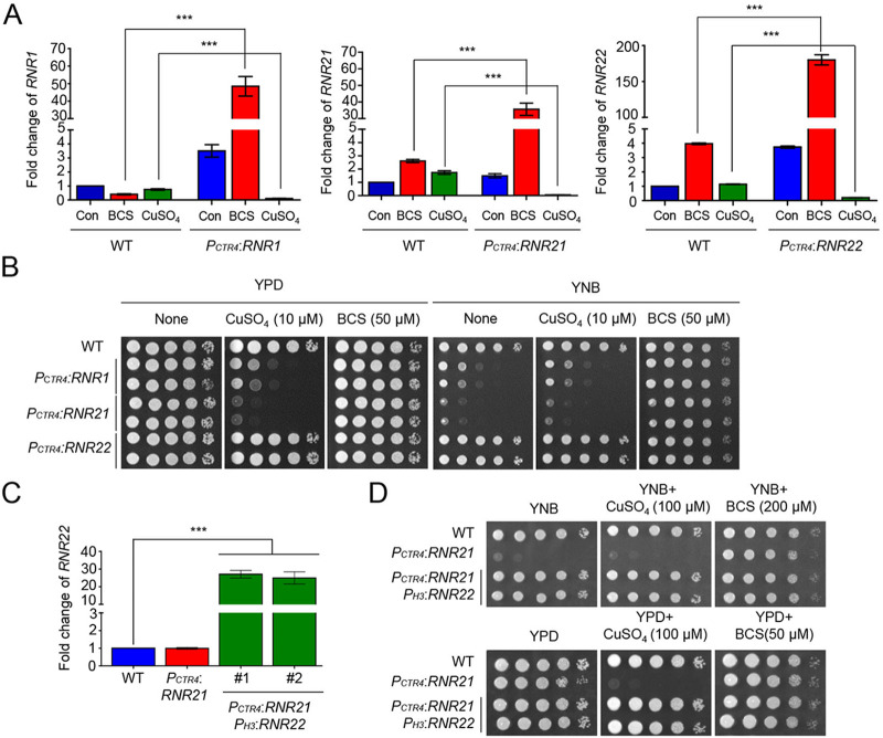
A balance in the deoxyribonucleotide (dNTPs) intracellular concentration is critical for the DNA replication and repair processes. In the model yeast Saccharomyces cerevisiae, the Mec1-Rad53-Dun1 kinase cascade mainly regulates the ribonucleotide reductase (RNR) gene expression during DNA replication and DNA damage stress. However, the RNR regulatory mechanisms in basidiomycete fungi during DNA replication and damage stress remain elusive. Here, we observed that in C. neoformans, RNR1 (large RNR subunit) and RNR21 (one small RNR subunit) were required for cell viability, but not RNR22 (another small RNR subunit). RNR22 overexpression compensated for the lethality of RNR21 suppression. In contrast to the regulatory mechanisms of RNRs in S. cerevisiae, Rad53 and Chk1 kinases cooperatively or divergently controlled RNR1 and RNR21 expression under DNA damage and DNA replication stress. In particular, this study revealed that Chk1 mainly regulated RNR1 expression during DNA replication stress, whereas Rad53, rather than Chk1, played a significant role in controlling the expression of RNR21 during DNA damage stress. Furthermore, the expression of RNR22, not but RNR1 and RNR21, was suppressed by the Ssn6-Tup1 complex during DNA replication stress. Notably, we observed that RNR1 expression was mainly regulated by Mbs1, whereas RNR21 expression was cooperatively controlled by Mbs1 and Bdr1 as downstream factors of Rad53 and Chk1 during DNA replication and damage stress. Collectively, the regulation of RNRs in C. neoformans has both evolutionarily conserved and divergent features in DNA replication and DNA damage stress, compared with other yeasts. IMPORTANCE Upon DNA replication or damage stresses, it is critical to provide proper levels of deoxynucleotide triphosphates (dNTPs) and activate DNA repair machinery. Ribonucleotide reductases (RNRs), which are composed of large and small subunits, are required for synthesizing dNTP. An imbalance in the intracellular concentration of dNTPs caused by the perturbation of RNR results in a reduction in DNA repair fidelity. Despite the importance of their roles, functions and regulations of RNR have not been elucidated in the basidiomycete fungi. In this study, we found that the roles of RNR1, RNR21, and RNR22 genes encoding RNR subunits in the viability of C. neoformans. Furthermore, their expression levels are divergently regulated by the Rad53-Chk1 pathway and the Ssn6-Tup1 complex in response to DNA replication and damage stresses. Therefore, this study provides insight into the regulatory mechanisms of RNR genes to DNA replication and damage stresses in basidiomycete fungi.
Keywords: Cryptococcus neoformans; DNA damage stress; DNA replication stress; ribonucleotide reductase.
Conflict of interest statement
The authors declare no conflict of interest.
Figures


Similar articles
-
Ixr1 is required for the expression of the ribonucleotide reductase Rnr1 and maintenance of dNTP pools.PLoS Genet. 2011 May;7(5):e1002061. doi: 10.1371/journal.pgen.1002061. Epub 2011 May 5. PLoS Genet. 2011. PMID: 21573136 Free PMC article.
-
Rad53- and Chk1-Dependent DNA Damage Response Pathways Cooperatively Promote Fungal Pathogenesis and Modulate Antifungal Drug Susceptibility.mBio. 2019 Jan 2;10(1):e01726-18. doi: 10.1128/mBio.01726-18. mBio. 2019. PMID: 30602579 Free PMC article.
-
Identification of RNR4, encoding a second essential small subunit of ribonucleotide reductase in Saccharomyces cerevisiae.Mol Cell Biol. 1997 Oct;17(10):6105-13. doi: 10.1128/MCB.17.10.6105. Mol Cell Biol. 1997. PMID: 9315670 Free PMC article.
-
DNA damage and cell cycle regulation of ribonucleotide reductase.Bioessays. 1993 May;15(5):333-9. doi: 10.1002/bies.950150507. Bioessays. 1993. PMID: 8343143 Review.
-
Function and regulation of yeast ribonucleotide reductase: cell cycle, genotoxic stress, and iron bioavailability.Biomed J. 2013 Mar-Apr;36(2):51-8. doi: 10.4103/2319-4170.110398. Biomed J. 2013. PMID: 23644233 Review.
Cited by
-
AoChk1 Is Required for Sporulation, Trap Formation, and Metabolic Process in Arthrobotrys oligospora.J Fungi (Basel). 2025 Aug 19;11(8):602. doi: 10.3390/jof11080602. J Fungi (Basel). 2025. PMID: 40863555 Free PMC article.
-
Identification of key transcription factors, including DAL80 and CRZ1, involved in heat and ethanol tolerance in Saccharomyces cerevisiae.Biotechnol Biofuels Bioprod. 2025 May 3;18(1):50. doi: 10.1186/s13068-025-02653-2. Biotechnol Biofuels Bioprod. 2025. PMID: 40319324 Free PMC article.
-
Pleiotropic roles of LAMMER kinase, Lkh1 in stress responses and virulence of Cryptococcus neoformans.Front Cell Infect Microbiol. 2024 May 7;14:1369301. doi: 10.3389/fcimb.2024.1369301. eCollection 2024. Front Cell Infect Microbiol. 2024. PMID: 38774630 Free PMC article.
-
Functional Characterization of DNA N-Glycosylase Ogg1 and Ntg1 in DNA Damage Stress of Cryptococcus neoformans.J Microbiol. 2023 Nov;61(11):981-992. doi: 10.1007/s12275-023-00092-y. Epub 2023 Dec 6. J Microbiol. 2023. PMID: 38055144
References
Publication types
MeSH terms
Substances
LinkOut - more resources
Full Text Sources
Miscellaneous
