

Extracellular Vesicles as Novel Drug-Delivery Systems through Intracellular Communications
- PMID: 35736256
- PMCID: PMC9230693
- DOI: 10.3390/membranes12060550
Extracellular Vesicles as Novel Drug-Delivery Systems through Intracellular Communications
Abstract
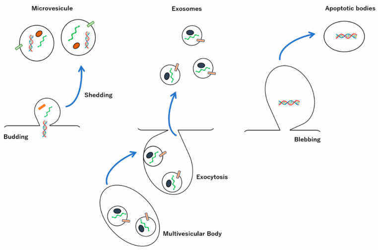
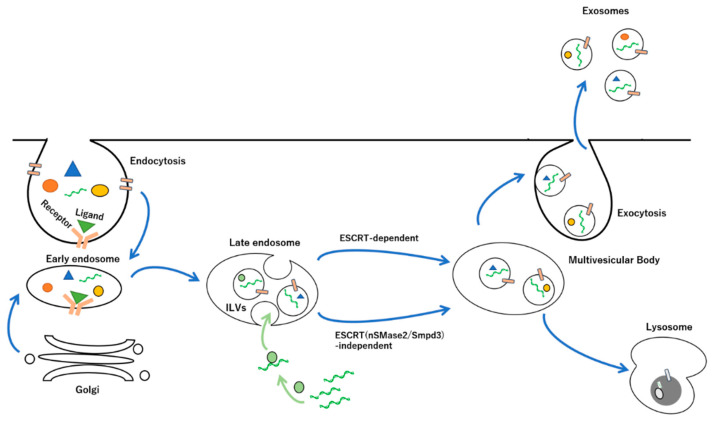
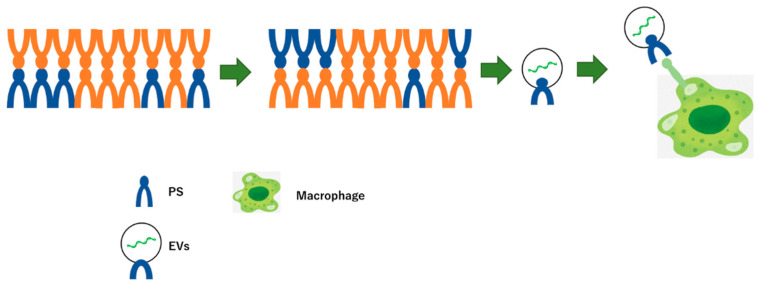
Since it has been reported that extracellular vesicles (EVs) carry cargo using cell-to-cell comminication according to various in vivo situations, they are exprected to be applied as new drug-delivery systems (DDSs). In addition, non-coding RNAs, such as microRNAs (miRNAs), have attracted much attention as potential biomarkers in the encapsulated extracellular-vesicle (EV) form. EVs are bilayer-based lipids with heterogeneous populations of varying sizes and compositions. The EV-mediated transport of contents, which includes proteins, lipids, and nucleic acids, has attracted attention as a DDS through intracellular communication. Many reports have been made on the development of methods for introducing molecules into EVs and efficient methods for introducing them into target vesicles. In this review, we outline the possible molecular mechanisms by which miRNAs in exosomes participate in the post-transcriptional regulation of signaling pathways via cell-cell communication as novel DDSs, especially small EVs.
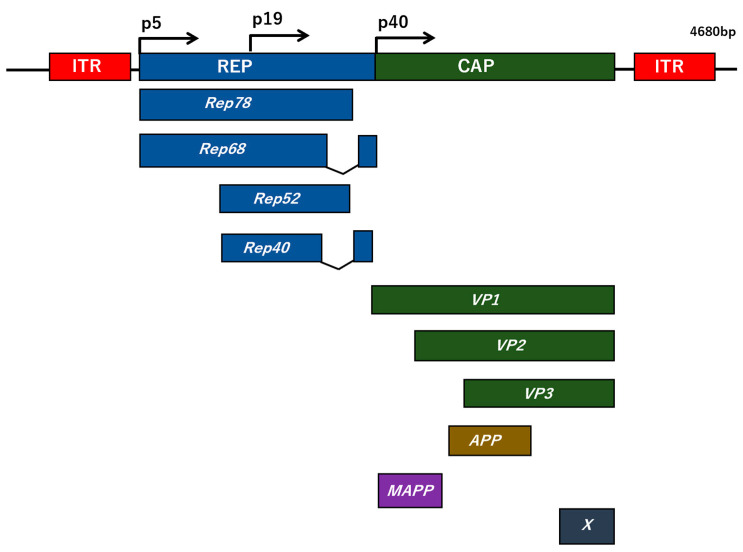
Keywords: adeno-associated virus; extracellular vesicles; intracellular communication; membrane vesicle production; non-coding RNAs.
Conflict of interest statement
The authors declare no conflict of interest.
Figures
Similar articles
-
Extracellular vesicle-based therapeutics: Extracellular vesicles as therapeutic targets and agents.Pharmacol Ther. 2023 Feb;242:108352. doi: 10.1016/j.pharmthera.2023.108352. Epub 2023 Jan 23. Pharmacol Ther. 2023. PMID: 36702209 Review.
-
Tumor-derived extracellular vesicles: reliable tools for Cancer diagnosis and clinical applications.Cell Commun Signal. 2019 Jul 10;17(1):73. doi: 10.1186/s12964-019-0390-y. Cell Commun Signal. 2019. PMID: 31291956 Free PMC article. Review.
-
Production of Extracellular Vesicles Loaded with Therapeutic Cargo.Methods Mol Biol. 2018;1831:37-47. doi: 10.1007/978-1-4939-8661-3_4. Methods Mol Biol. 2018. PMID: 30051423 Free PMC article.
-
Delivery of microRNAs by Extracellular Vesicles in Viral Infections: Could the News be Packaged?Cells. 2019 Jun 18;8(6):611. doi: 10.3390/cells8060611. Cells. 2019. PMID: 31216738 Free PMC article. Review.
-
Role of lipid signalling in extracellular vesicles-mediated cell-to-cell communication.Cytokine Growth Factor Rev. 2023 Oct;73:20-26. doi: 10.1016/j.cytogfr.2023.08.006. Epub 2023 Aug 23. Cytokine Growth Factor Rev. 2023. PMID: 37648617 Review.
Cited by
-
Anti-Inflammatory Effects of Helminth-Derived Products: Potential Applications and Challenges in Diabetes Mellitus Management.J Inflamm Res. 2024 Dec 28;17:11789-11812. doi: 10.2147/JIR.S493374. eCollection 2024. J Inflamm Res. 2024. PMID: 39749005 Free PMC article. Review.
-
CAR-T-Derived Extracellular Vesicles: A Promising Development of CAR-T Anti-Tumor Therapy.Cancers (Basel). 2023 Feb 7;15(4):1052. doi: 10.3390/cancers15041052. Cancers (Basel). 2023. PMID: 36831396 Free PMC article. Review.
-
The Fatal Role of Enterohaemorrhagic Escherichia coli Shiga Toxin-associated Extracellular Vesicles in Host Cells.J Microbiol. 2023 Aug;61(8):715-727. doi: 10.1007/s12275-023-00066-0. Epub 2023 Sep 4. J Microbiol. 2023. PMID: 37665555 Review.
-
BMP-7 modified exosomes derived from synovial mesenchymal stem cells attenuate osteoarthritis by M2 polarization of macrophages.Heliyon. 2023 Sep 10;9(9):e19934. doi: 10.1016/j.heliyon.2023.e19934. eCollection 2023 Sep. Heliyon. 2023. PMID: 37809369 Free PMC article.
-
Molecular Docking and Intracellular Translocation of Extracellular Vesicles for Efficient Drug Delivery.Int J Mol Sci. 2022 Oct 26;23(21):12971. doi: 10.3390/ijms232112971. Int J Mol Sci. 2022. PMID: 36361760 Free PMC article. Review.
References
Publication types
Grants and funding
LinkOut - more resources
Full Text Sources
