Review
. 2022 May 31;12(6):500.
doi: 10.3390/metabo12060500.

Metabolic Determinants in Cardiomyocyte Function and Heart Regenerative Strategies

Affiliations
Review

Metabolic Determinants in Cardiomyocyte Function and Heart Regenerative Strategies

Magda Correia et al. Metabolites. .

Abstract

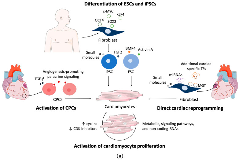
Heart disease is the leading cause of mortality in developed countries. The associated pathology is characterized by a loss of cardiomyocytes that leads, eventually, to heart failure. In this context, several cardiac regenerative strategies have been developed, but they still lack clinical effectiveness. The mammalian neonatal heart is capable of substantial regeneration following injury, but this capacity is lost at postnatal stages when cardiomyocytes become terminally differentiated and transit to the fetal metabolic switch. Cardiomyocytes are metabolically versatile cells capable of using an array of fuel sources, and the metabolism of cardiomyocytes suffers extended reprogramming after injury. Apart from energetic sources, metabolites are emerging regulators of epigenetic programs driving cell pluripotency and differentiation. Thus, understanding the metabolic determinants that regulate cardiomyocyte maturation and function is key for unlocking future metabolic interventions for cardiac regeneration. In this review, we will discuss the emerging role of metabolism and nutrient signaling in cardiomyocyte function and repair, as well as whether exploiting this axis could potentiate current cellular regenerative strategies for the mammalian heart.

Keywords: cardiac regeneration; cardiomyocytes; cell reprogramming; metabolism; mitochondria; nutrient signaling; pluripotency.


Conflict of interest statement

The authors declare no conflict of interest.

Figures

Figure 1
Figure 1
Connection between cardiomyocyte development and mitochondria usage and maturation, highlighting mitochondria specialization according to the spacial arrangement in postnatal cardiomyocytes. After birth, a bioenergetic shift from glucose-driven anaerobic glycolysis to the oxidation of fuels in the mitochondria occurs. Mitochondria increase in size and density, and cristae become denser. The upregulation of PGC coactivators and the downregulation of HIF1α regulate mitochondria biogenesis. Abbreviations: FAO, fatty acid oxidation; HIF, hypoxia-inducible factor; IMF, intermyofibrillar; OXPHOS, oxidative phosphorylation; PGC, peroxisome proliferator-activated receptor-gamma coactivator; PN, perinuclear mitochondria; SS, subsarcolemmal; TCA, tricarboxylic acid cycle; up arrow: increase; low arrow: decrease.
Figure 2
Figure 2
Energetic metabolism of cardiomyocytes. FFAs (free fatty acids), the main energetic fuel, are converted into long-chain acyl-CoA esters by fatty acyl CoA synthetase, and then, into long-chain acylcarnitine by CPT1, the rate limiting enzyme of long-chain FAO. Inside the mitochondria, long-chain acylcarnitine is converted back to long-chain acyl-CoA by CPT2, and then, several isoforms of acyl-CoA dehydrogenase, enoyl-CoA hydratase, 3-hydroxyacyl CoA dehydrogenase, and 3-ketoacyl CoA thiolase (specific for different chain lengths) mediate the shortening of the fatty acyl moiety, generating acetyl-CoA, FADH2, and NADH. Glc is transported by GLUTs (GLUT4 in adult heart) into cardiomyocytes, and, in the cytosol, Glc is converted into pyruvate through glycolysis. Pyruvate may be oxidized in mitochondria or converted into lactate by LDH in the cytosol. Lactate uptake and secretion is mediated by MCTs (MCT4 in the adult heart). MCT1 makes part of the mtLOC, which is also composed of CD147, LDH, and COX. Lactate may be oxidized in the mitochondria by this complex. Ketones, mostly βOHB, may feed TCA after being metabolized by the enzymes β-hydroxybutyrate dehydrogenase 1 (BDH1, the enzyme that interconverts β-hydroxybutyrate into acetoacetate) and succinyl-CoA:3-ketoacid CoA transferase (SCOT, the rate-limiting enzyme of ketone oxidation). SCFAs (including acetate, propanoate, and butyrate) can cross the mitochondrial membrane by diffusion, and enter FAO through SCAD, after being activated via ACSMs. BCAA and other amino acids, such as Glu, may be used for energetic purposes by feeding the TCA cycle. BCATm and BCKDH catalyze the first two enzymatic steps in BCAA metabolism. The contribution of these metabolic pathways changes in the injured heart. In ischemia (Isc), the reliance on glucose and lactate increases, but when insulin signaling is repressed, FAO is upregulated. During heart failure (HF) glucose oxidation prevails until mitochondria functionality is impaired, which happens at advanced stages of disease. In diabetes mellitus (DM), glucose uptake and oxidation decrease, inducing a higher reliance of cardiac metabolism on lipids for energetic purposes. Abbreviations: α-KG, α-ketoglutarate; βOHB, β-hydroxybutyrate; AA, amino acid; AcAc, acetoacetate; ACSMs, acyl-coenzyme A synthetase medium chain family members; AGE, advanced glycation end products; Alb, albumin; ATP, adenosine-5’-triphosphate; BCAA, branched chain amino acids; BCATm, mitochondrial branched-chain aminotransferase; BCKDH, branched-chain α-ketoacid dehydrogenase complex; BDH1, 3-hydroxybutyrate dehydrogenase 1; CD147, cluster of differentiation 147; COX, cytochrome c oxidase; CPT, carnitine palmitoyl transferase; DM, diabetes mellitus; FA-carnitine, acyl-carnitine; FA-CoA, long-chain acyl-CoA; FABP, fatty acid binding protein; FAO, fatty acid oxidation; FAT/CD36, fatty acid translocase; FATP, fatty acid transport protein; FADH2, reduced flavin adenine dinucleotide; GDH, glutamate dehydrogenase; Glc, glucose; Gln, glutamine; Glu, glutamate; GLUT, glucose transporter; GluT, glutamate transporter; GlyS, glycogen synthesis; GS, glutamine synthase; HBP, hexosamine biosynthetic pathway; HF, heart failure; Isc, ischemia; LD, lipid droplet; LDH, lactate dehydrogenase; MCT, monocarboxylate transporter; MPC, mitochondrial pyruvate carrier; NAD+, oxidized nicotinamide adenine dinucleotide; NADH, reduced nicotinamide adenine dinucleotide; O-GlcNAc, O-linked β-N-acetylglucosamine; OXPHOS, oxidative phosphorylation; PDH, pyruvate dehydrogenase; PDK, pyruvate dehydrogenase kinase; PPP, pentose phosphate pathway; Pyr, pyruvate; R-CoA, branched-chain acyl CoAs; SCOT, succinyl-CoA: 3-oxoacid-CoA transferase; SCAD, short chain acyl-CoA dehydrogenase; SCFA, short chain fatty acids; SLC16A, solute carrier family 16 member 1; Suc-CoA, succinyl-CoA; TCA, tricarboxylic acid cycle.
Figure 3
Figure 3
Heart regeneration strategies and metabolic manipulations to increase cardiac regeneration. (a) Approaches for replacing lost cardiomyocytes. By modulating factors that either promote (overexpression of cyclins) or repress (silencing of CDK inhibitors) cell cycle activity, adult cardiomyocytes, can be forced to proliferate. Metabolic and signaling pathways, as well as the expression of non-coding RNAs, can also be modulated to induce cardiomyocyte proliferation. Stimulating the differentiation of CPCs into cardiomyocytes can be achieved by treatment with TGF-β or paracrine signaling, for example. ESCs and iPSCs have been used to generate cardiomyocytes, which can be transplanted to replace cells that were lost upon cardiac injury. Direct cardiac reprogramming can be achieved by overexpressing the cardiac factors Mef2c, Gata4, and Tbx5 (termed MGT) into resident fibroblasts. The addition of additional transcription factors and small molecules, and manipulation of miRNAs, can improve reprograming efficiency. (b) Metabolic reprogramming to increase cardiac regenerative potential. The stimulation of glycolysis, inhibition of FAO, and decrease in oxygen increase cardiomyocyte proliferation. Activation of CPCs can be induced with high concentrations of glucose and glutamine in the media, hypoxia, and reduction in ROS levels. Differentiation of ESCs and iPSCs into cardiomyocytes can be improved by increasing FAO, decreasing glucose, and increasing exogenous lipids and galactose. For direct cardiac reprogramming, stimulation of OXPHOS and inhibition of glycolysis increases the efficiency of the process. Abbreviations: CPCs, cardiac progenitor cells; ESCs, embryonic stem cells; FAO, fatty acid oxidation; iPSCs, induced pluripotent stem cells; OXPHOS, oxidative phosphorylation; ROS, reactive oxygen species; TCA, Tricarboxylic acid cycle; up arrow: increase; low arrow: decrease.
Figure 3
Figure 3
Heart regeneration strategies and metabolic manipulations to increase cardiac regeneration. (a) Approaches for replacing lost cardiomyocytes. By modulating factors that either promote (overexpression of cyclins) or repress (silencing of CDK inhibitors) cell cycle activity, adult cardiomyocytes, can be forced to proliferate. Metabolic and signaling pathways, as well as the expression of non-coding RNAs, can also be modulated to induce cardiomyocyte proliferation. Stimulating the differentiation of CPCs into cardiomyocytes can be achieved by treatment with TGF-β or paracrine signaling, for example. ESCs and iPSCs have been used to generate cardiomyocytes, which can be transplanted to replace cells that were lost upon cardiac injury. Direct cardiac reprogramming can be achieved by overexpressing the cardiac factors Mef2c, Gata4, and Tbx5 (termed MGT) into resident fibroblasts. The addition of additional transcription factors and small molecules, and manipulation of miRNAs, can improve reprograming efficiency. (b) Metabolic reprogramming to increase cardiac regenerative potential. The stimulation of glycolysis, inhibition of FAO, and decrease in oxygen increase cardiomyocyte proliferation. Activation of CPCs can be induced with high concentrations of glucose and glutamine in the media, hypoxia, and reduction in ROS levels. Differentiation of ESCs and iPSCs into cardiomyocytes can be improved by increasing FAO, decreasing glucose, and increasing exogenous lipids and galactose. For direct cardiac reprogramming, stimulation of OXPHOS and inhibition of glycolysis increases the efficiency of the process. Abbreviations: CPCs, cardiac progenitor cells; ESCs, embryonic stem cells; FAO, fatty acid oxidation; iPSCs, induced pluripotent stem cells; OXPHOS, oxidative phosphorylation; ROS, reactive oxygen species; TCA, Tricarboxylic acid cycle; up arrow: increase; low arrow: decrease.

Similar articles

Cited by

References

    1. Virani S.S., Alonso A., Aparicio H.J., Benjamin E.J., Bittencourt M.S., Callaway C.W., Carson A.P., Chamberlain A.M., Cheng S., Delling F.N., et al. Heart Disease and Stroke Statistics 2021 Update. Circulation. 2021;143:e254–e743. doi: 10.1161/CIR.0000000000000950. - DOI - PubMed
    1. Laflamme M.A., Murry C.E. Heart regeneration. Nature. 2011;473:326–335. doi: 10.1038/nature10147. - DOI - PMC - PubMed
    1. Hashimoto H., Olson E.N., Bassel-Duby R. Therapeutic approaches for cardiac regeneration and repair. Nat. Rev. Cardiol. 2018;15:585–600. doi: 10.1038/s41569-018-0036-6. - DOI - PMC - PubMed
    1. Ghiroldi A., Piccoli M., Cirillo F., Monasky M.M., Ciconte G., Pappone C., Anastasia L. Cell-Based Therapies for Cardiac Regeneration: A Comprehensive Review of Past and Ongoing Strategies. Int. J. Mol. Sci. 2018;19:3194. doi: 10.3390/ijms19103194. - DOI - PMC - PubMed
    1. Neidig L.E., Weinberger F., Palpant N.J., Mignone J., Martinson A.M., Sorensen D.W., Bender I., Nemoto N., Reinecke H., Pabon L., et al. Evidence for minimal cardiogenic potential of stem cell antigen 1-positive cells in the adult mouse heart. Circulation. 2018;138:2960–2962. doi: 10.1161/CIRCULATIONAHA.118.035273. - DOI - PMC - PubMed

LinkOut - more resources