

Hesperetin, a Citrus Flavonoid, Ameliorates Inflammatory Cytokine-Mediated Inhibition of Oligodendroglial Cell Morphological Differentiation
- PMID: 35736620
- PMCID: PMC9230394
- DOI: 10.3390/neurolint14020039
Hesperetin, a Citrus Flavonoid, Ameliorates Inflammatory Cytokine-Mediated Inhibition of Oligodendroglial Cell Morphological Differentiation
Abstract
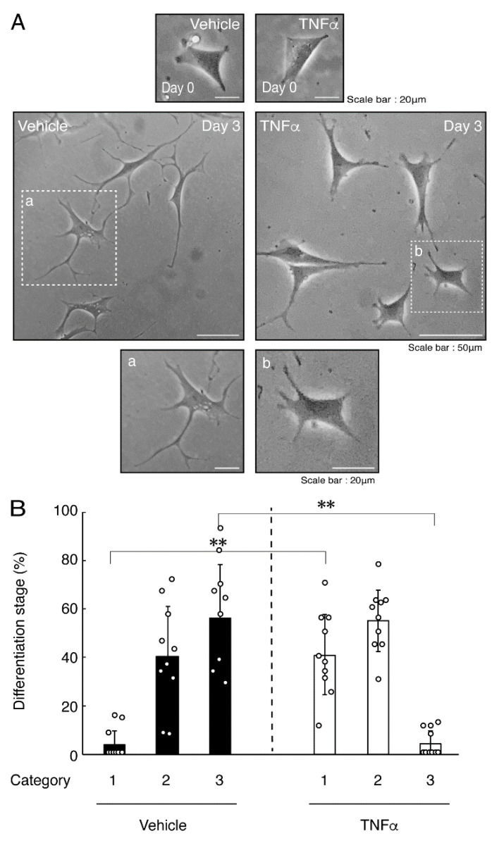
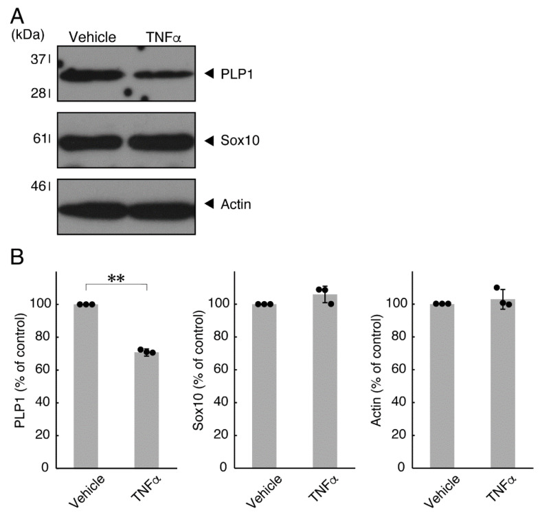
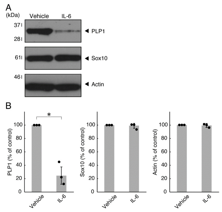
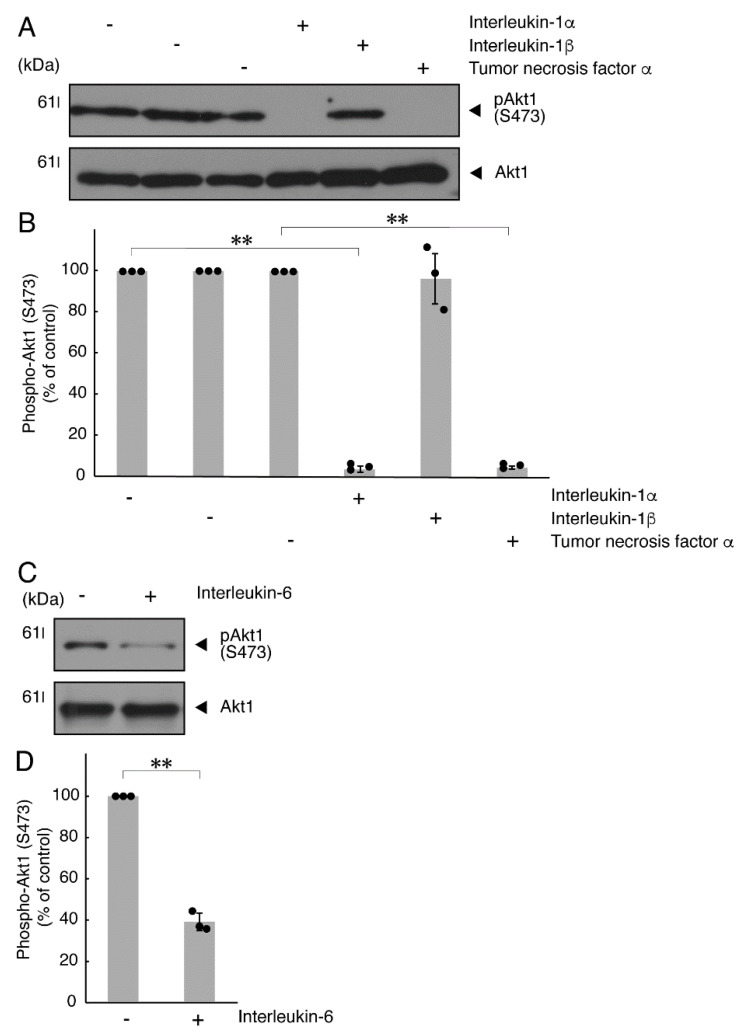
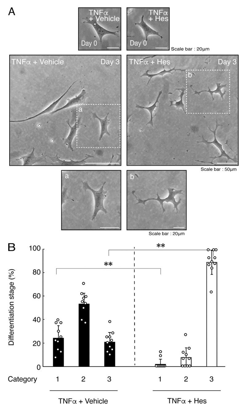
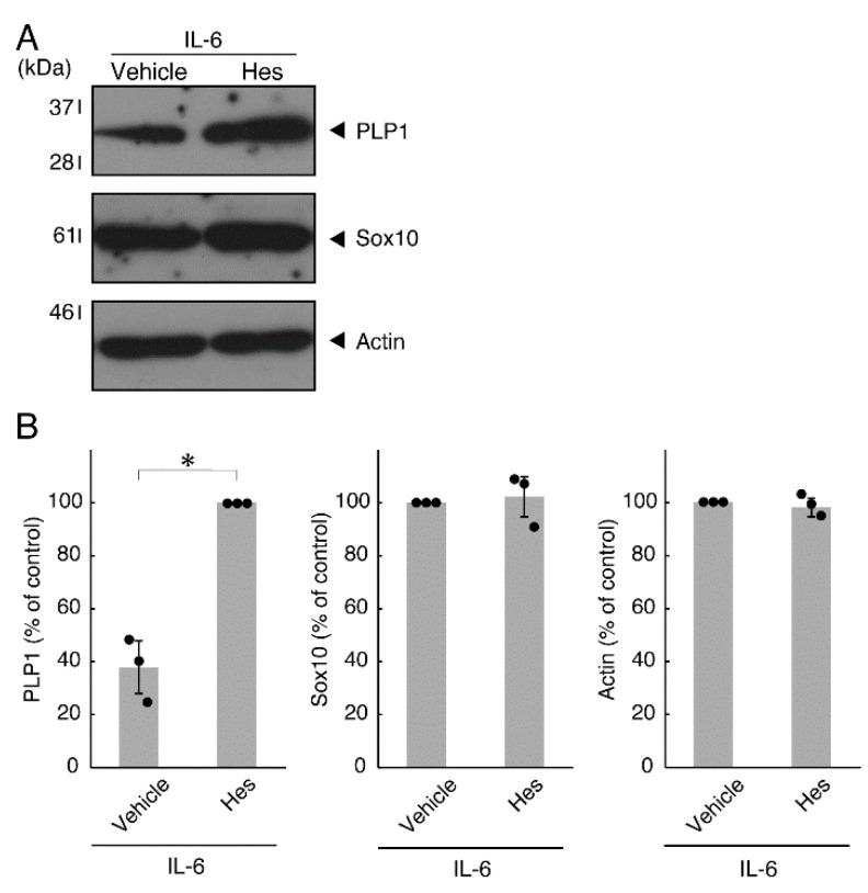
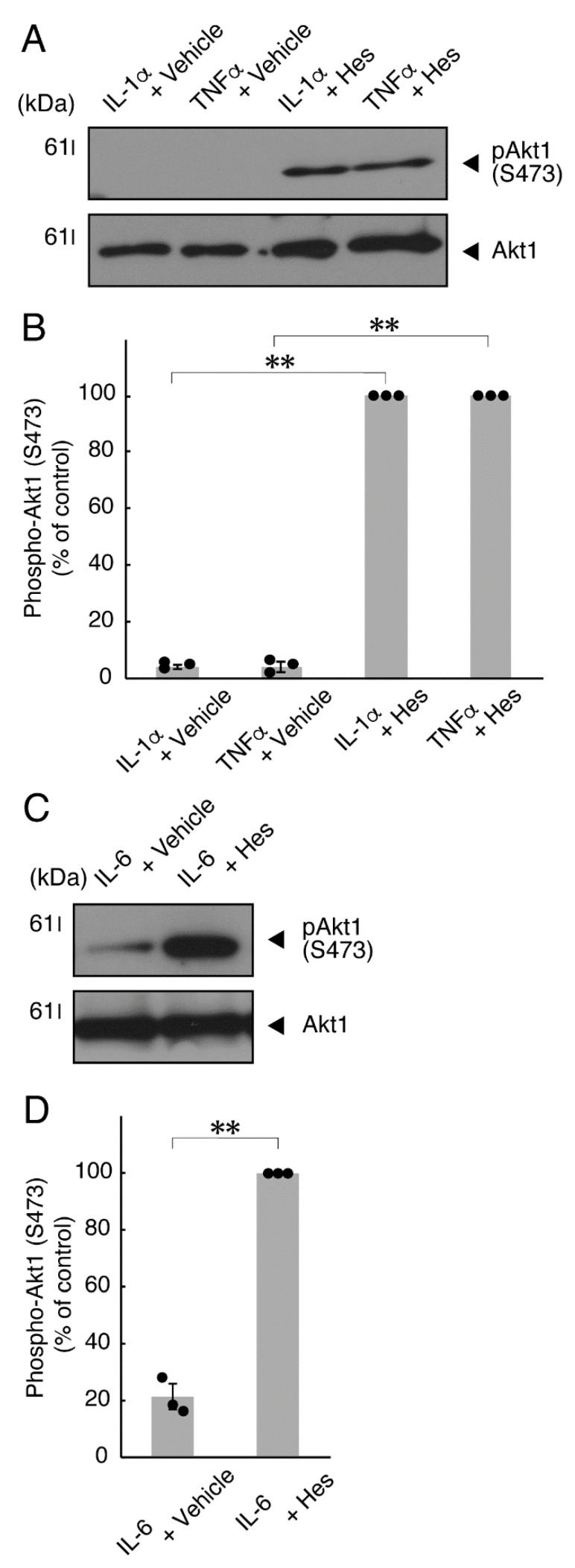
Oligodendrocytes (oligodendroglial cells) are glial cells that wrap neuronal axons with their differentiated plasma membranes called myelin membranes. In the pathogenesis of inflammatory cytokine-related oligodendroglial cell and myelin diseases such as multiple sclerosis (MS), typical inflammatory cytokines tumor necrosis factor α (TNFα) and interleukin-6 (IL-6) are thought to contribute to the degeneration and/or progression of the degeneration of oligodendroglial cells and, in turn, the degeneration of naked neuronal cells in the central nervous system (CNS) tissues. Despite the known involvement of these inflammatory cytokines in disease progression, it has remained unclear whether and how TNFα or IL-6 affects the oligodendroglial cells themselves or indirectly. Here we show that TNFα or IL-6 directly inhibits morphological differentiation in FBD-102b cells, which are differentiation models of oligodendroglial cells. Their phenotype changes were supported by the decreased expression levels of oligodendroglial cell differentiation and myelin marker proteins. In addition, TNFα or IL-6 decreased phosphorylation levels of Akt kinase, whose upregulation has been associated with promoting oligodendroglial cell differentiation. Hesperetin, a flavonoid mainly contained in citrus fruit, is known to have neuroprotective effects. Hesperetin might also be able to resolve pre-illness conditions, including the irregulated secretion of cytokines, through diet. Notably, the addition of hesperetin into cells recovered TNFα- or IL-6-induced inhibition of differentiation, as supported by increased levels of marker protein expression and phosphorylation of Akt kinase. These results suggest that TNFα or IL-6 itself contributes to the inhibitory effects on the morphological differentiation of oligodendroglial cells, possibly providing information not only on their underlying pathological effects but also on flavonoids with potential therapeutic effects at the molecular and cellular levels.
Keywords: Akt kinase; IL-6; TNFα; differentiation; hesperetin; oligodendrocyte.
Conflict of interest statement
The authors have declared that no competing interests exist.
Figures


Similar articles
-
Knockdown of Rab7B, But Not of Rab7A, Which Antagonistically Regulates Oligodendroglial Cell Morphological Differentiation, Recovers Tunicamycin-Induced Defective Differentiation in FBD-102b Cells.J Mol Neurosci. 2023 Jun;73(6):363-374. doi: 10.1007/s12031-023-02117-y. Epub 2023 May 30. J Mol Neurosci. 2023. PMID: 37248316
-
Hyaluronic acid and its receptor CD44, acting through TMEM2, inhibit morphological differentiation in oligodendroglial cells.Biochem Biophys Res Commun. 2022 Oct 8;624:102-111. doi: 10.1016/j.bbrc.2022.07.092. Epub 2022 Jul 31. Biochem Biophys Res Commun. 2022. PMID: 35940122
-
Hesperetin Ameliorates Inhibition of Neuronal and Oligodendroglial Cell Differentiation Phenotypes Induced by Knockdown of Rab2b, an Autism Spectrum Disorder-Associated Gene Product.Neurol Int. 2023 Mar 10;15(1):371-391. doi: 10.3390/neurolint15010025. Neurol Int. 2023. PMID: 36976668 Free PMC article.
-
Neuronal injury in chronic CNS inflammation.Best Pract Res Clin Anaesthesiol. 2010 Dec;24(4):551-62. doi: 10.1016/j.bpa.2010.11.001. Epub 2010 Nov 29. Best Pract Res Clin Anaesthesiol. 2010. PMID: 21619866 Review.
-
Defective Oligodendroglial Lineage and Demyelination in Amyotrophic Lateral Sclerosis.Int J Mol Sci. 2021 Mar 26;22(7):3426. doi: 10.3390/ijms22073426. Int J Mol Sci. 2021. PMID: 33810425 Free PMC article. Review.
Cited by
-
Investigating the Protective Effects of a Citrus Flavonoid on the Retardation Morphogenesis of the Oligodendroglia-like Cell Line by Rnd2 Knockdown.Neurol Int. 2023 Dec 26;16(1):33-61. doi: 10.3390/neurolint16010003. Neurol Int. 2023. PMID: 38251051 Free PMC article.
-
Hesperetin-7-O-glucoside/β-cyclodextrin Inclusion Complex Induces Acute Vasodilator Effect to Inhibit the Cold Sensation Response during Localized Cold-Stimulate Stress in Healthy Human Subjects: A Randomized, Double-Blind, Crossover, and Placebo-Controlled Study.Nutrients. 2023 Aug 24;15(17):3702. doi: 10.3390/nu15173702. Nutrients. 2023. PMID: 37686734 Free PMC article. Clinical Trial.
-
Hypomyelinating Leukodystrophy 10 (HLD10)-Associated Mutations of PYCR2 Form Large Size Mitochondria, Inhibiting Oligodendroglial Cell Morphological Differentiation.Neurol Int. 2022 Dec 16;14(4):1062-1080. doi: 10.3390/neurolint14040085. Neurol Int. 2022. PMID: 36548190 Free PMC article.
-
Rehabilitation treatment of multiple sclerosis.Front Immunol. 2023 Apr 6;14:1168821. doi: 10.3389/fimmu.2023.1168821. eCollection 2023. Front Immunol. 2023. PMID: 37090712 Free PMC article. Review.
-
Knockdown of Rab7B, But Not of Rab7A, Which Antagonistically Regulates Oligodendroglial Cell Morphological Differentiation, Recovers Tunicamycin-Induced Defective Differentiation in FBD-102b Cells.J Mol Neurosci. 2023 Jun;73(6):363-374. doi: 10.1007/s12031-023-02117-y. Epub 2023 May 30. J Mol Neurosci. 2023. PMID: 37248316
References
LinkOut - more resources
Full Text Sources
