Review
. 2022 Jun 3;29(2):243-280.
doi: 10.3390/pathophysiology29020020.

The Role of Exposomes in the Pathophysiology of Autoimmune Diseases II: Pathogens

Affiliations
Review

The Role of Exposomes in the Pathophysiology of Autoimmune Diseases II: Pathogens

Aristo Vojdani et al. Pathophysiology. .

Abstract

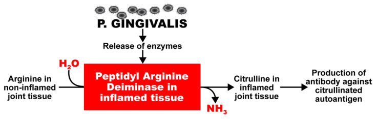
In our continuing examination of the role of exposomes in autoimmune disease, we use this review to focus on pathogens. Infections are major contributors to the pathophysiology of autoimmune diseases through various mechanisms, foremost being molecular mimicry, when the structural similarity between the pathogen and a human tissue antigen leads to autoimmune reactivity and even autoimmune disease. The three best examples of this are oral pathogens, SARS-CoV-2, and the herpesviruses. Oral pathogens reach the gut, disturb the microbiota, increase gut permeability, cause local inflammation, and generate autoantigens, leading to systemic inflammation, multiple autoimmune reactivities, and systemic autoimmunity. The COVID-19 pandemic put the spotlight on SARS-CoV-2, which has been called "the autoimmune virus." We explore in detail the evidence supporting this. We also describe how viruses, in particular herpesviruses, have a role in the induction of many different autoimmune diseases, detailing the various mechanisms involved. Lastly, we discuss the microbiome and the beneficial microbiota that populate it. We look at the role of the gut microbiome in autoimmune disorders, because of its role in regulating the immune system. Dysbiosis of the microbiota in the gut microbiome can lead to multiple autoimmune disorders. We conclude that understanding the precise roles and relationships shared by all these factors that comprise the exposome and identifying early events and root causes of these disorders can help us to develop more targeted therapeutic protocols for the management of this worldwide epidemic of autoimmunity.

Keywords: autoantibodies; autoimmunity; bystander activation; environmental trigger; epitope spreading; exposome; infection; molecular mimicry; pathogen; pathophysiology.


Conflict of interest statement

A.V. is co-owner, CEO, and technical director of Immunosciences Lab., Inc. E.V. is the owner and founder of Regenera Medical. A.Z.R. and Y.S. declare no conflict of interest.

Figures

Figure 1
Figure 1
Potentiation by oral pathogens. Oral pathogens such as P. gingivalis can potentiate the deamination of arginine or formation of citrullinated proteins and peptides in joint and other tissues.
Figure 2
Figure 2
Mechanism by which oral infection can lead to multiple autoimmune reactivities. Oral infectious agents reach the gut and disturb the gut flora, leading to a disrupted epithelium and gut permeability. Local inflammation generates autoantigens by PAD; antibody production starts against one autoantigen, but through epitope spreading reaches out to multiple antigens, leading to systemic inflammation, multiple autoimmune reactivities, and systemic autoimmunity.
Figure 3
Figure 3
The interactive network of the immune system. The immune system is a complex network of different kinds of cells that must work together and combine in just the right numbers, ratios, and percentages to protect the host from pathogens.
Figure 4
Figure 4
Peptide molecular mimicry between SARS-CoV-2 spike protein and mitochondria M2, F-actin, and thyroid peroxidase (TPO) [36].
Figure 5
Figure 5
Diagram of possible relationship between SARS-CoV-2 proteins and autoimmune target proteins.
Figure 6
Figure 6
Elevations in IgG antibody against 27 different human tissue antigens in the blood of patients with mild to severe SARS-CoV-2 infection (positive for PCR and antibody) in comparison to healthy controls.
Figure 7
Figure 7
Elevations in IgA antibody against 29 different human tissue antigens in the blood of patients with mild to severe SARS-CoV-2 infection (positive for PCR and antibody) in comparison to healthy controls.
Figure 8
Figure 8
Epstein–Barr virus and a few of its associated autoimmune diseases.
Figure 9
Figure 9
HHV-6 and a few of its associated autoimmune diseases.
Figure 10
Figure 10
10 key pathological processes induced by the herpes family of viruses that results in the destruction of neurons and the production of antibodies against MBP, MOG, and other neuronal antigens.
Figure 11
Figure 11
How viruses can induce autoimmunity through molecular mimicry. Viruses are taken up by APCs and broken up into viral antigens that are structurally similar to self-antigen. When the APC primes the T cell with the viral antigen, it becomes an autoreactive T cell and attacks both viruses and self-tissue.
Figure 12
Figure 12
How viruses can induce autoimmunity through bystander activation. Excessive non-specific antiviral immune responses lead to the release of inflammatory cytokines and self-antigens. Self-antigens are presented to autoreactive T cells, which attack the innocent “bystander” tissues.
Figure 13
Figure 13
How viruses can induce autoimmunity through erroneous bystander activation by disturbed immune surveillance. The body’s defender cells usually constantly monitor and actively protect the body through immune surveillance, calling upon the proper defensive immune cells when a threat is recognized. However, a disturbance of the immune surveillance can lead to the recruitment of autoreactive Th1 and Th1/Th17 cells instead, which can result in tissue destruction.
Figure 14
Figure 14
How viruses can induce autoimmunity through epitope spreading. Persistent viral infection and continued tissue damage leads to the release of even more and more new self-antigens, increasing the number of self-tissue targets as the autoreactive T cells spread their attacks to more epitopes.
Figure 15
Figure 15
Proposed mechanisms by which microbiome dysbiosis may induce autoimmune disease. Environmental factors such as toxic chemicals, food antigens, and pathogens can affect mucosal inflammation, intestinal barrier function, and immunity, causing increased translocation of bacteria or their metabolites, such as circulating endotoxin LPS. This promotes systemic aberrant anti-inflammatory responses, eventually resulting in autoimmune disorders.
Figure 16
Figure 16
Exposome factors and early events that are involved in the breach of barriers and immune defenses that are associated with inflammatory, autoimmune, neurodegenerative disorders, allergies, and hypersensitivities.

Similar articles

Cited by

References

    1. Vojdani A., Vojdani E. The Role of Exposomes in the Pathophysiology of Autoimmune Diseases I: Toxic Chemicals and Food. Pathophysiology. 2021;28:513–543. doi: 10.3390/pathophysiology28040034. - DOI - PMC - PubMed
    1. Wild C.P. Complementing the genome with an “exposome”: The outstanding challenge of environmental exposure measurement in molecular epidemiology. Cancer Epidemiol. Biomarkers Prev. 2005;14:1847–1850. doi: 10.1158/1055-9965.EPI-05-0456. - DOI - PubMed
    1. Vineis P., Barouki R. The exposome as the science of social-to-biological transitions. Environ. Int. 2022;165 doi: 10.1016/j.envint.2022.107312. - DOI - PubMed
    1. Kristo A., Klimis-Zacas D., Sikalidis A. Protective Role of Dietary Berries in Cancer. Antioxidants. 2016;5:37. doi: 10.3390/antiox5040037. - DOI - PMC - PubMed
    1. Sikalidis A.K. Amino Acids and Immune Response: A Role for Cysteine, Glutamine, Phenylalanine, Tryptophan and Arginine in T-cell Function and Cancer? Pathol. Oncol. Res. 2015;21:9–17. doi: 10.1007/s12253-014-9860-0. - DOI - PubMed

LinkOut - more resources