. 2022 Jun 1;10(2):19.
doi: 10.3390/proteomes10020019.

A Comparison of Blood Plasma Small Extracellular Vesicle Enrichment Strategies for Proteomic Analysis

Affiliations

A Comparison of Blood Plasma Small Extracellular Vesicle Enrichment Strategies for Proteomic Analysis

Natalie P Turner et al. Proteomes. .

Abstract

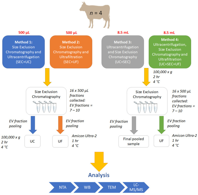
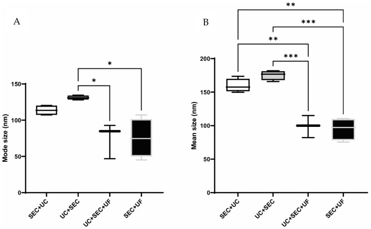
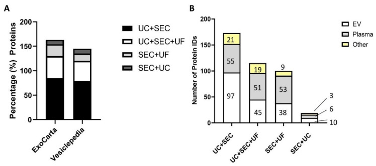
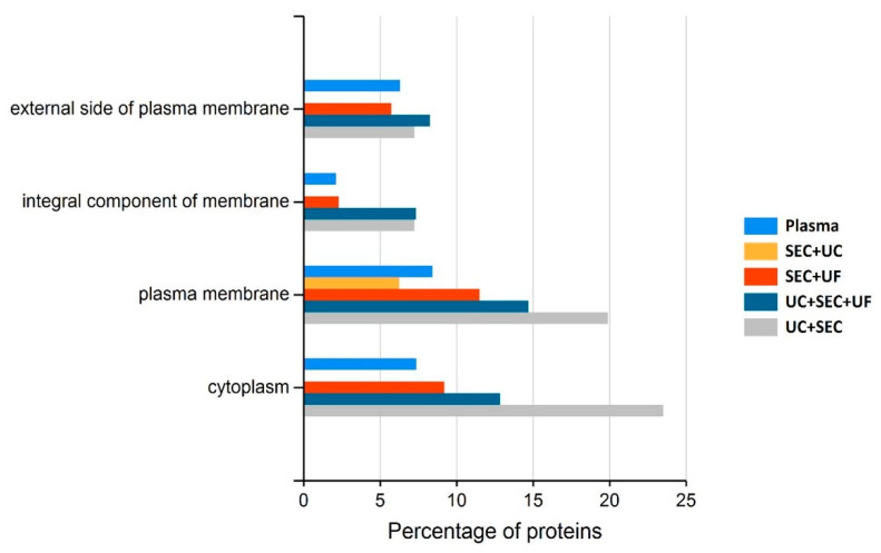
Proteomic analysis of small extracellular vesicles (sEVs) poses a significant challenge. A 'gold-standard' method for plasma sEV enrichment for downstream proteomic analysis is yet to be established. Methods were evaluated for their capacity to successfully isolate and enrich sEVs from plasma, minimise the presence of highly abundant plasma proteins, and result in the optimum representation of sEV proteins by liquid chromatography tandem mass spectrometry. Plasma from four cattle (Bos taurus) of similar physical attributes and genetics were used. Three methods of sEV enrichment were utilised: ultracentrifugation (UC), size-exclusion chromatography (SEC), and ultrafiltration (UF). These methods were combined to create four groups for methodological evaluation: UC + SEC, UC + SEC + UF, SEC + UC and SEC + UF. The UC + SEC method yielded the highest number of protein identifications (IDs). The SEC + UC method reduced plasma protein IDs compared to the other methods, but also resulted in the lowest number of protein IDs overall. The UC + SEC + UF method decreased sEV protein ID, particle number, mean and mode particle size, particle yield, and did not improve purity compared to the UC + SEC method. In this study, the UC + SEC method was the best method for sEV protein ID, purity, and overall particle yield. Our data suggest that the method and sequence of sEV enrichment strategy impacts protein ID, which may influence the outcome of biomarker discovery studies.

Keywords: enrichment; exosome; extracellular vesicle; extracellular vesicles; isolation; mass spectrometry; proteomics; size-exclusion chromatography; small extracellular vesicle; ultracentrifugation; ultrafiltration.


Conflict of interest statement

The authors declare no conflict of interest.

Figures

Figure 1
Figure 1
Workflow for small extracellular vesicle (sEV) enrichment methods 1–4. SEC: size-exclusion chromatography; UF: ultrafiltration; UC: ultracentrifugation; EV: extracellular vesicle; NTA: nanoparticle tracking analysis; WB: western blot; TEM: transmission electron microscopy; LC-MS/MS: liquid chromatography tandem mass spectrometry.
Figure 2
Figure 2
Western blot of bovine serum albumin (BSA) in SEC (A) and UC + SEC (B) fractions. L = ladder; C = Control, 1 ug purified BSA; B = blank; 7, 8, 9, 10, 11, 12, 13, 14, 15, 16, 1–6 = individual sEV (7–10), non-sEV (11–16) fractions, and void volume fractions pool (1–6). Predicted molecular weight of BSA = 69 kDa. (Full length blot images available in Supplementary File S2—Figures S1 and S2).
Figure 3
Figure 3
Mode (A) and mean (B) size distributions of pooled sEV fractions by all methods. In both mean and mode size analysis, UC + SEC particles were significantly larger than those obtained by the UC + SEC + UF and SEC + UF methods (UC + SEC + UF, n = 3; all other methods, n = 4; error bars ± SEM; * p < 0.05, ** p < 0.01, *** p < 0.001). The mean size of particles obtained by the SEC + UC method were significantly larger than the UC + SEC + UF and SEC + UF methods.
Figure 4
Figure 4
(A) Percentage of top 100 EV proteins (Vesiclepedia and ExoCarta) identified in sEV enriched samples by all methods. (B) Summary plot of mapped EV and non-EV proteins in the four methods under study identified in the Vesiclepedia complete database and plasma control. EV proteins = identified in samples and Vesiclepedia database; Plasma = identified in plasma and sEV-enriched sample; Other = identified in sEV-enriched sample only.
Figure 5
Figure 5
Functional enrichment analysis (cellular component) of detected proteins in plasma processed using one of the four methods for sEV protein enrichment and plasma control.

Similar articles

Cited by

References

    1. Collins B.C., Hunter C.L., Liu Y., Schilling B., Rosenberger G., Bader S.L., Chan D.W., Gibson B.W., Gingras A.-C., Held J.M., et al. Multi-laboratory assessment of reproducibility, qualitative and quantitative performance of SWATH-mass spectrometry. Nat. Commun. 2017;8:291. doi: 10.1038/s41467-017-00249-5. - DOI - PMC - PubMed
    1. Wiśniewski J.R., Zougman A., Nagaraj N., Mann M. Universal sample preparation method for proteome analysis. Nat. Methods. 2009;6:359–362. doi: 10.1038/nmeth.1322. - DOI - PubMed
    1. Record M., Carayon K., Poirot M., Silvente-Poirot S. Exosomes as new vesicular lipid transporters involved in cell–cell communication and various pathophysiologies. Biochim. et Biophys. Acta (BBA)-Mol. Cell Biol. Lipids. 2014;1841:108–120. doi: 10.1016/j.bbalip.2013.10.004. - DOI - PubMed
    1. Zhang Y., Liu Y., Liu H., Tang W.H. Exosomes: Biogenesis, biologic function and clinical potential. Cell Biosci. 2019;9:19. doi: 10.1186/s13578-019-0282-2. - DOI - PMC - PubMed
    1. Nazimek K., Bryniarski K., Santocki M., Ptak W. Exosomes as Mediators of Intercellular Communication: Clinical Implications. Pol. Arch. Intern. Med. 2015;125:370–380. doi: 10.20452/pamw.2840. - DOI - PubMed