. 2022 Jul 5;94(26):9345-9354.
doi: 10.1021/acs.analchem.2c00905. Epub 2022 Jun 23.

Human 2D Crypt Model for Assaying Intestinal Stem Cell Proliferation and Differentiation

Affiliations

Human 2D Crypt Model for Assaying Intestinal Stem Cell Proliferation and Differentiation

Yuli Wang et al. Anal Chem. .

Abstract

Intestine is a common site of adverse drug effects in clinical trials; thus, improved in vitro models for preclinical screening of pharmaceutical compounds are sought. A planar, self-renewing human intestinal monolayer platform based on primary adult gastrointestinal stem cells, termed the 2D crypt model, has been developed to screen for the effects of various compounds on the intestinal epithelium. The 2D crypt platform is based on a standard 12-well plate format and consists of cell culture inserts with a collagen film overlaying an impermeable film patterned with an array of micron-scale holes. This two-chamber format enables a gradient of growth factors to be applied such that the tissue self-organizes into spatially segregated stem and differentiated cell compartments. The patterned monolayer mimics a gut epithelium in possessing a stem cell niche, migrating proliferative and differentiated cells. Once established, the 2D crypts replicate many aspects of in vivo physiology, including cell migration, maturation, and apoptotic cell death. The planar geometry of the system simplifies dosing, sampling, and imaging during assay. An immunofluorescence-based assay was established to quantitatively assess cell density, proliferation, migration, viability, and the abundance and localization of postmitotic lineages as a function of time. The model was used to perform a small-scale screen of compounds, including signaling molecules, endogenous hormones/cytokines, and microbial metabolites, on tissue homeostasis. Hit compounds that significantly impacted proliferation and/or differentiation were readily identified. The 2D crypt platform represents a significant innovation in the development of microphysiological systems for emulating the gut epithelium for compound screens.


Conflict of interest statement

Y.W., C.E.S., and N.L.A. disclose a financial interest in Altis Biosystems, Inc.

Figures

Figure 1.
Figure 1.
Establishment of a 2D crypt array possessing patterned zones of proliferative and differentiated cells. (A) Schematic of the platform and crypt formation. (B-G) Immunofluorescence staining of primary human colon epithelial cell growth on the 2D crypt microdevice after 5 days polarization under DM/EM. (B) From left to right: staining of proliferative cells (EdU, green), goblet cells (Muc2, yellow), colonocytes (ALP, red), and merged images. (C) Staining of enteroendocrine cells (ChgA, red). Inserted image: high magnification of an enteroendocrine cell. (D) Proliferative cells were stained with Ki67. (E) Staining of tight junction ZO-1 (red). (F) Staining of adherens junction β-catenin (red). (G) Staining of proliferative cells (EdU, green) and differentiated cells (KRT-20, purple). Top: low magnification image shows 9×9 crypt units are uniformly polarized. Inserted is the high magnification image showing one crypt unit. Bottom: XZ cross-sectional image showing the upward protrusion of the proliferative zone. DNA in nuclei were stained with Hoechst 33342 (blue). Transverse colon cells from donor-1 were used. Scale=200 μm unless specified.
Figure 2.
Figure 2.
Optimization of the collagen layer thickness and the center-to-center gap of microholes. (A) Schematic of the attributes. (B) Top row: EdU (green) and Hoechst 33342 (blue) staining of 2D crypts (microhole diameter=70 μm, gap=350 μm) on collagen layer of different thicknesses. Bottom: Side view of a cross-section through a fluorescein-labelled collagen scaffold. (C) EdU (green), Muc2 (yellow), ALP (red) and Hoechst 33342 (blue) staining of 2D crypts with different center-to-center gaps (all are 70 μm diameter microholes). (D) Quantification of DNA, EdU, Muc2 and ALP coverage. Coverage % is defined in experimental section. Polarization duration=5 days. n=3 arrays, t-test. **: p<0.005; *: p<0.05. Transverse colon cells from donor-1 were used.
Figure 3.
Figure 3.
Effect of differentiation duration (0–11 days) on expression of EdU, Muc2 and ALP in human 2D crypts. (A) EdU (green), Muc2 (yellow), ALP (red) and Hoechst 33342 (blue) staining of 2D crypts at varying times after the switch to EM/DM (basal/luminal medium). (B-D) Quantification of EdU, Muc2 and ALP coverage per crypt unit. n=3 arrays. Coverage % is defined in experimental section. Transverse colon cells from donor-1 were used.
Figure 4.
Figure 4.
Self-renewal, cell migration and apoptosis in 2D crypts. (A) Cell migration was revealed by an EdU pulse chase experiment. At 4 days after polarization, the cells were pulsed with EdU for 24 h. Post EdU pulse, the cells were fixed at 0–4 days and the incorporated EdU developed with a fluorescent reagent. The proliferative zone of one crypt unit is marked with dotted circle. (B) Radius of EdU+ zone vs. post-EdU pulse duration. n=30 crypt units for each measurement. (C) Brightfield image after crypt polarization for 5 and 10 days. (D) Detection of apoptotic cells (annexin-V, green) and necrotic cells (PI, red). (E) Detection of apoptotic cells (caspase-3, red) and proliferative cells (EdU, green). (F) Percentage of active caspase-3 coverage (average and standard deviation) in the proliferative and differentiated cell zones. n=3 arrays. Coverage % is defined in experimental section. Center-to-center gap is 525 μm for (A) and (B), 350 μm for (C)-(F). Transverse colon cells from donor-1 were used.
Figure 5.
Figure 5.
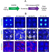
Impact of compounds on cell proliferation and differentiation. (A) Schematic of the assay steps. (B) Representative images of 4 unit crypts exposed to the indicated compounds. The 2D crypts were from transverse colon of donor-1. Green: EdU, yellow: Muc2, red: ALP, blue: DNA. Transverse colon cells from donor-1 were used.
Figure 6.
Figure 6.
Screening of 12 compounds on cell proliferation and differentiation. (A-C) Relative fluorescence intensity of 10×10 crypt units (normalized to the control untreated crypts and total cell count). n=3. *: p<0.05; **: p<0.005. (A) Proliferative cells (EdU+). (B) Goblet cells (Muc2+). (C) Colonocytes cells (ALP+). (D) List of 12 compounds, the selected concentration, references for the concentration, and magnitude of effect. Transverse colon cells from donor-1 and donor-2 were used.

References

    1. Koh A; De Vadder F; Kovatcheva-Datchary P; Bäckhed F From dietary fiber to host physiology: short-chain fatty acids as key bacterial metabolites. Cell 2016, 165, 1332–1345. - PubMed
    1. Andrews C; McLean MH; Durum SK Cytokine Tuning of Intestinal Epithelial Function. Front. Immunol 2018, 9, 1270. - PMC - PubMed
    1. Chen Y; Lin YN; Davis KM; Wang QR; Rnjak-Kovacina J; Li CM; Isberg RR; Kumamoto CA; Mecsas J; Kaplan DL Robust bioengineered 3D functional human intestinal epithelium. Sci. Rep 2015, 5, 13708. - PMC - PubMed
    1. Sung JH; Yu J; Luo D; Shuler ML; March JC Microscale 3-D hydrogel scaffold for biomimetic gastrointestinal (GI) tract model. Lab Chip 2011, 11, 389–392. - PubMed
    1. Meunier V; Bourrié M; Berger Y; Fabre G The human intestinal epithelial cell line Caco-2; pharmacological and pharmacokinetic applications. Cell Biol. Toxicol 1995, 11, 187–194. - PubMed

Publication types