. 2022 Aug;23(8):e13705.
doi: 10.1002/acm2.13705. Epub 2022 Jun 23.

A standardized workflow for respiratory-gated motion management decision-making

Affiliations

A standardized workflow for respiratory-gated motion management decision-making

Sandra M Meyers et al. J Appl Clin Med Phys. 2022 Aug.

Abstract

Purpose: Motion management of tumors within the lung and abdomen is challenging because it requires balancing tissue sparing with accuracy of hitting the target, while considering treatment delivery efficiency. Physicists can play an important role in analyzing four-dimensional computed tomography (4DCT) data to recommend the optimal respiratory gating parameters for a patient. The goal of this work was to develop a standardized procedure for making recommendations regarding gating parameters and planning margins for lung and gastrointestinal stereotactic body radiotherapy (SBRT) treatments. In doing so, we hoped to simplify decision-making and analysis, and provide a tool for troubleshooting complex cases.

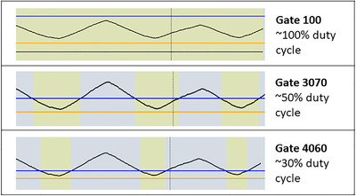
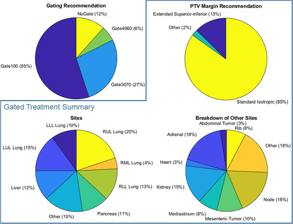
Methods: Factors that impact gating decisions and planning target volume (PTV) margins were identified. The gating options included gating on exhale with approximately a 50% duty cycle (Gate3070), exhale gating with a reduced duty cycle (Gate4060), and treating for most of respiration, excluding only extreme inhales and exhales (Gate100). A standard operating procedure was developed, as well as a physics consult document to communicate motion management recommendations to other members of the treatment team. This procedure was implemented clinically for 1 year and results are reported below.

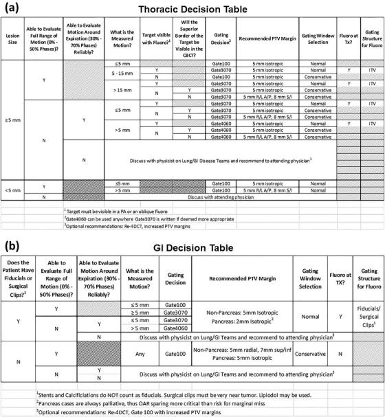
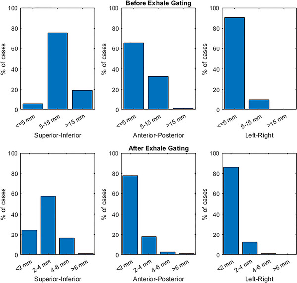
Results: Identified factors that impact motion management included the magnitude of motion observed on 4DCT, the regularity of breathing and quality of 4DCT data, and ability to observe the target on fluoroscopy. These were collated into two decision tables-one specific to lung tumors and another for gastrointestinal tumors-such that a physicist could answer a series of questions to determine the optimal gating and PTV margin. The procedure was used clinically for 252 sites from 213 patients treated with respiratory-gated SBRT and standardized practice across our 12-member physics team.

Conclusion: Implementation of a standardized procedure for respiratory gating had a positive impact in our clinic, improving efficiency and ease of 4DCT analysis and standardizing gating decision-making amongst physicists.

Keywords: gastrointestinal cancer; lung cancer; motion management; respiratory gating; stereotactic body radiotherapy.


Conflict of interest statement

Kelly Kisling, Todd Atwood, and Xenia Ray acknowledge Honoraria and speaker fees from Varian Medical Systems. Xenia Ray has a research agreement with Varian Medical Systems.

Figures

FIGURE 1
FIGURE 1
Examples of relatively normal respiratory signals from Varian's Real‐time Position ManagementTM (RPM) system (Varian Medical Systems, Palo Alto, CA) during treatment delivery, where a high amplitude corresponds to inhale (0% phase), and a low amplitude corresponds to exhale (50% phase). The gating window is defined by the user by adjusting the location of the blue and orange lines, and beam‐on is indicated by yellow. Three potential gating schemes are shown, along with their corresponding gating window: (1 ‐ top) the Gate 100 window is set to encompass all regular phases of breathing motion; (2 ‐ middle) the Gate 3070 window approximately encompasses phases 30% to 70%, and thus the beam is on approximately half the time; (3 ‐ bottom) Gate 4060 encompasses approximately 40% to 60% phases, so the beam is on roughly one third of the time
FIGURE 2
FIGURE 2
Depiction of a tumor position in each phase of the breathing cycle and corresponding amplitude of the respiratory trace
FIGURE 3
FIGURE 3
Example of a lung tumor that can be visualized in fluoroscopy with an oblique projection angle. (a) Axial, coronal, and sagittal average CT images, demonstrating that the tumor is positioned posterior to the heart, which will obscure visualization in anterior–posterior projection fluoroscopy. (a) also depicts the beam's eye view of an oblique angled fluoroscopic imaging field, where separation can be observed between the heart (red) and ITV (orange) structures. (b) The ITV and PTV contours (green) and the tumor position on both (b) inhale and (c) exhale oblique fluoroscopy. The corresponding amplitudes of the breathing trace for these images are shown by the dotted green lines in (d). Using fluoroscopy, the gating window (orange and blue lines) is set so that beam‐on (indicated by yellow colorwash) will only occur while the tumor is inside the ITV. This patient is an example of a Gate4060 treatment
FIGURE 4
FIGURE 4
Motion management decision tables for thoracic (a) and GI (b) tumors. N, no; TX, treatment; Y, yes
FIGURE 5
FIGURE 5
Example cases where thoracic tumors are not visible in fluoroscopic imaging. (a) Sagittal CT image showing tumor located posterior to the diaphragm and inferior to the apex of the diaphragm, such that diaphragm will obscure tumor visualization in an anterior–posterior projection. (b) Axial CT image showing a diffuse tumor, or ground glass object, with signal that is too faint to be observed in fluoroscopy. (c) Axial, sagittal, and coronal CT images of a tumor that is located posterior to mediastinal soft tissue, such as the heart
FIGURE 6
FIGURE 6
Summary of patients for which physics 4D analysis was performed, where charts inside the blue box summarize SBRT cases that were treated with respiratory‐gated motion management (i.e., excluding “NoGate” cases). Standard isotropic PTV margins are 5 mm for lung and GI, and 2 mm for pancreas with implanted fiducials. Extended superior–inferior margins feature additional superior–inferior expansions on the standard isotropic margins of 7 mm for GI and 8 mm for lung. LLL, left lower lobe; LUL, left lower lobe; RLL, right lower lobe; RML, right medial lobe; RUL, right upper lobe
FIGURE 7
FIGURE 7
Motion of SBRT treatment targets in all three directions before gating (i.e., during free breathing) and after performing exhale respiratory gating (i.e., Gate3070 or Gate4060), as measured on the 4DCT and reported in the physics 4D consult form

References

    1. Prezzano KM, Ma SJ, Hermann GM, Rivers CI, Gomez‐Suescun JA, Singh AK. Stereotactic body radiation therapy for non‐small cell lung cancer: a review. World J Clin Oncol. 2019;10(1):14‐27. 10.5306/wjco.v10.i1.14 - DOI - PMC - PubMed
    1. Bockbrader M, Kim E. Role of intensity‐modulated radiation therapy in gastrointestinal cancer. Expert Rev Anticancer Ther. 2009;9(5):637‐647. 10.1586/era.09.16 - DOI - PubMed
    1. Rusthoven KE, Kavanagh BD, Cardenes H, et al. Multi‐institutional phase I/II trial of stereotactic body radiation therapy for liver metastases. J Clin Oncol Off J Am Soc Clin Oncol. 2009;27(10):1572‐1578. 10.1200/JCO.2008.19.6329 - DOI - PubMed
    1. Bujold A, Massey CA, Kim JJ, et al. Sequential phase I and II trials of stereotactic body radiotherapy for locally advanced hepatocellular carcinoma. J Clin Oncol Off J Am Soc Clin Oncol. 2013;31(13):1631‐1639. 10.1200/JCO.2012.44.1659 - DOI - PubMed
    1. Liu HH, Balter P, Tutt T, et al. Assessing respiration‐induced tumor motion and internal target volume using four‐dimensional computed tomography for radiotherapy of lung cancer. Int J Radiat Oncol Biol Phys. 2007;68(2):531‐540. 10.1016/j.ijrobp.2006.12.066 - DOI - PubMed