Review
. 2022 Aug:140:59-97.
doi: 10.1016/j.clinph.2022.04.022. Epub 2022 May 18.

Transcranial magnetic stimulation of the brain: What is stimulated? - A consensus and critical position paper

Affiliations
Review

Transcranial magnetic stimulation of the brain: What is stimulated? - A consensus and critical position paper

Hartwig R Siebner et al. Clin Neurophysiol. 2022 Aug.

Abstract

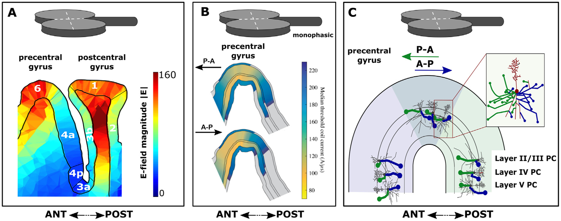
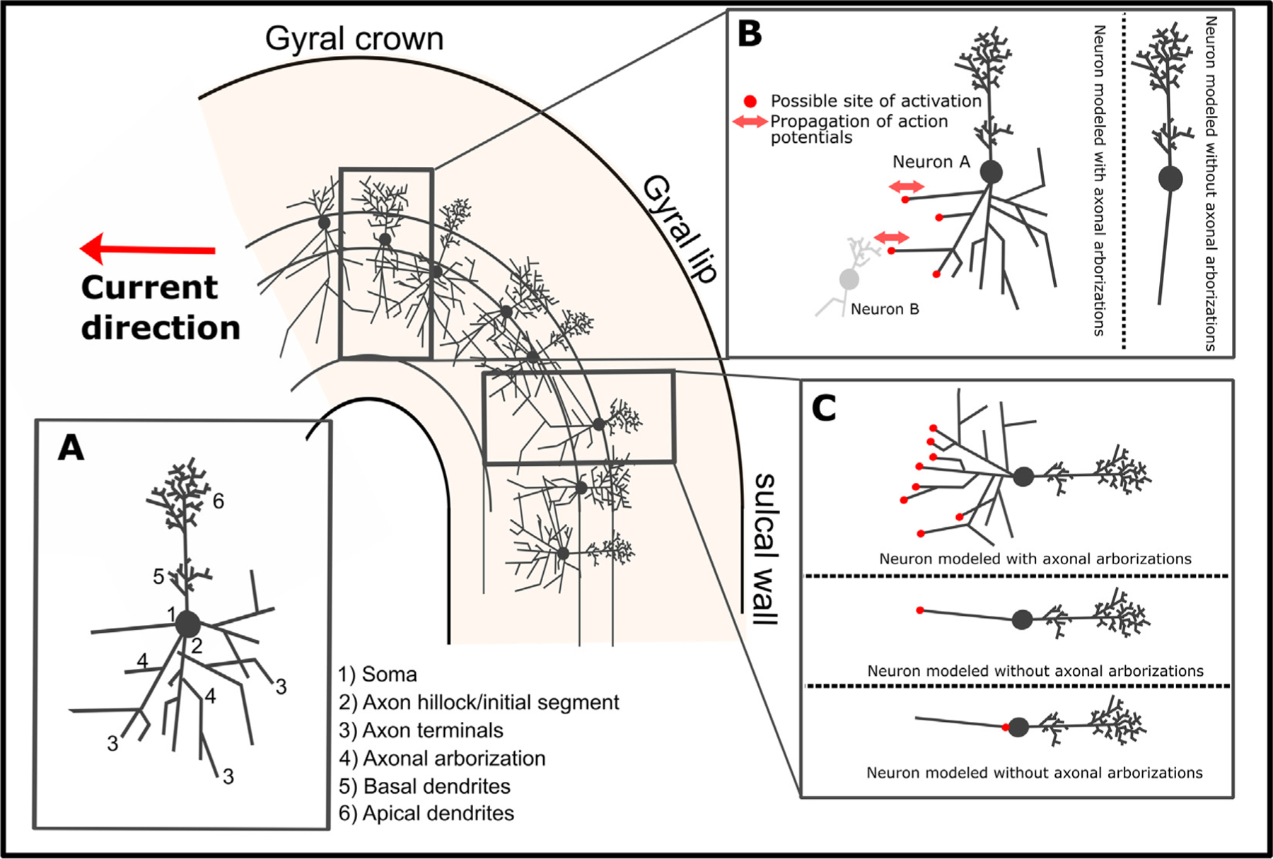
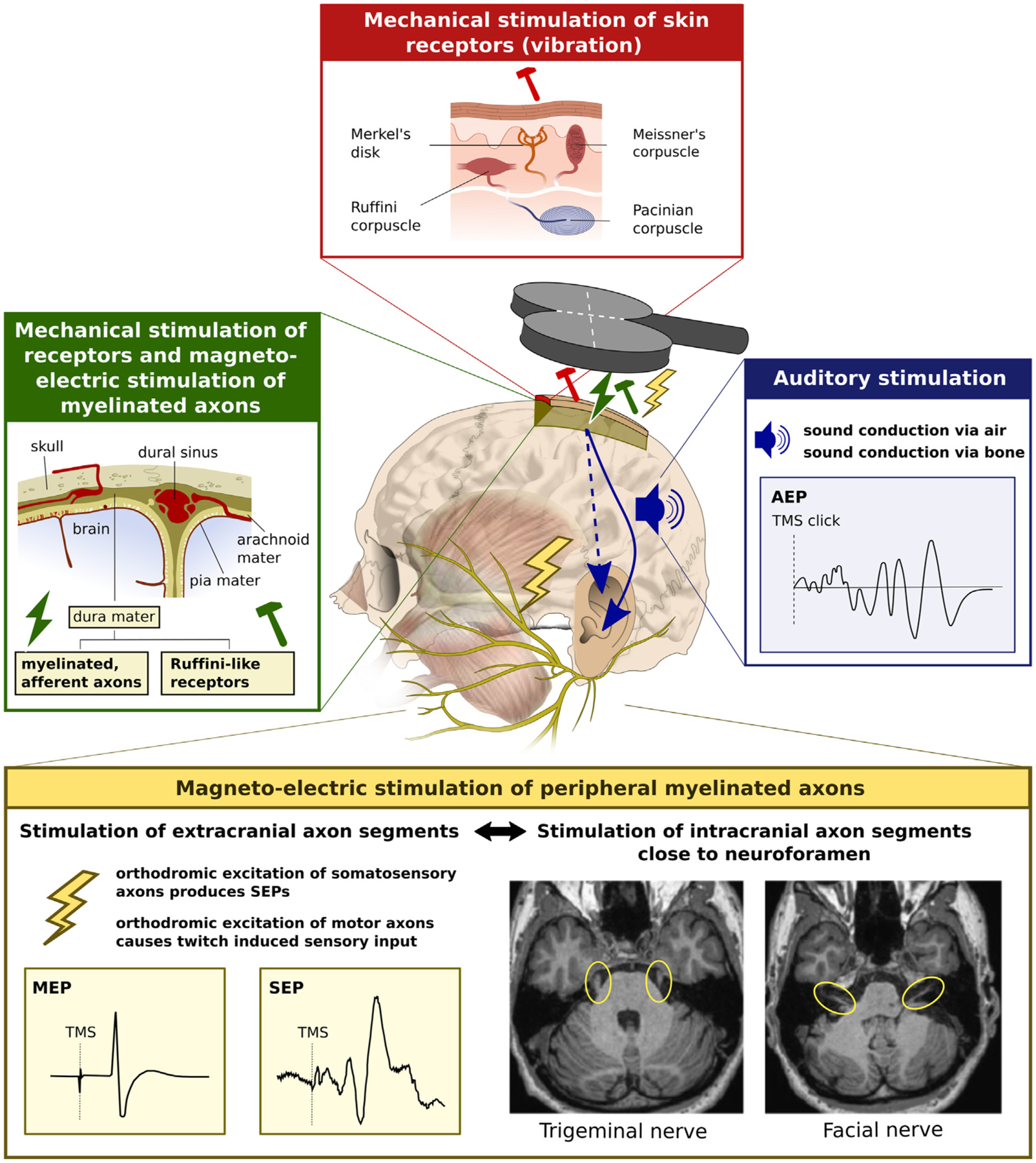
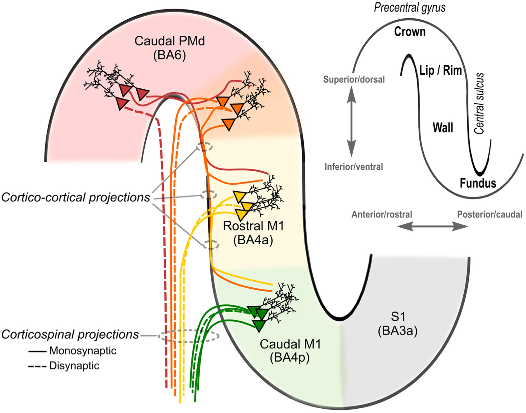
Transcranial (electro)magnetic stimulation (TMS) is currently the method of choice to non-invasively induce neural activity in the human brain. A single transcranial stimulus induces a time-varying electric field in the brain that may evoke action potentials in cortical neurons. The spatial relationship between the locally induced electric field and the stimulated neurons determines axonal depolarization. The induced electric field is influenced by the conductive properties of the tissue compartments and is strongest in the superficial parts of the targeted cortical gyri and underlying white matter. TMS likely targets axons of both excitatory and inhibitory neurons. The propensity of individual axons to fire an action potential in response to TMS depends on their geometry, myelination and spatial relation to the imposed electric field and the physiological state of the neuron. The latter is determined by its transsynaptic dendritic and somatic inputs, intrinsic membrane potential and firing rate. Modeling work suggests that the primary target of TMS is axonal terminals in the crown top and lip regions of cortical gyri. The induced electric field may additionally excite bends of myelinated axons in the juxtacortical white matter below the gyral crown. Neuronal excitation spreads ortho- and antidromically along the stimulated axons and causes secondary excitation of connected neuronal populations within local intracortical microcircuits in the target area. Axonal and transsynaptic spread of excitation also occurs along cortico-cortical and cortico-subcortical connections, impacting on neuronal activity in the targeted network. Both local and remote neural excitation depend critically on the functional state of the stimulated target area and network. TMS also causes substantial direct co-stimulation of the peripheral nervous system. Peripheral co-excitation propagates centrally in auditory and somatosensory networks, but also produces brain responses in other networks subserving multisensory integration, orienting or arousal. The complexity of the response to TMS warrants cautious interpretation of its physiological and behavioural consequences, and a deeper understanding of the mechanistic underpinnings of TMS will be critical for advancing it as a scientific and therapeutic tool.

Keywords: Mechanism of action; Motor cortex; Physiology; Transcranial magnetic stimulation.


Conflict of interest statement

Declaration of Competing Interest The authors declare the following financial interests/personal relationships which may be considered as potential competing interests: Andrea Antal has received honoraria from NeuroCare GmbH, Germany and Savir GmbH, Germany. Peter T. Fox has received patents in multiple jurisdictions for various TMS technologies, including the cortical column cosine model (C3) and the delivery of transcranial magnetic stimulation in accordance with the C3 model in an image-guided or robotically controlled manner. These patents are assigned to the University of Texas Board of Regents and have been licensed for commercialization by the University of Texas to a private entity in which Dr. Fox has ownership interest. Mark Hallett is an inventor of patents held by NIH for an immunotoxin for the treatment of focal movement disorders and the H-coil for magnetic stimulation; in relation to the latter, he has received license fee payments from the NIH (from Brainsway). He is on the Medical Advisory Boards of CALA Health and Brainsway (both unpaid positions). He is on the Editorial Board of approximately 15 journals and receives royalties and/or honoraria from publishing from Cambridge University Press, Oxford University Press, Springer, Wiley, Wolters Kluwer, and Elsevier. He has research grants from Medtronic, Inc. for a study of DBS for dystonia and CALA Health for studies of a device to suppress tremor. Hartwig R. Siebner has received honoraria as speaker from Sanofi Genzyme, Denmark and Novartis, Denmark, as consultant from Sanofi Genzyme, Denmark, Lophora, Denmark, and Lundbeck AS, Denmark, and as editor-in-chief (Neuroimage Clinical) and senior editor (NeuroImage) from Elsevier Publishers, Amsterdam, The Netherlands. He has received royalties as book editor from Springer Publishers, Stuttgart, Germany and from Gyldendal Publishers, Copenhagen, Denmark. Yoshikazu Ugawa has received honoraria from Takeda Pharmaceutical Company Limited, Eisai Co., Ltd., FP Pharmaceutical Corporation, Otsuka Pharmaceutical Co., Ltd., Elsevier Japan K. K., Kyowa Hakko Kirin Co., Ltd., Dainippon Sumitomo Pharma Co., Ltd., Mitsubishi Tanabe Pharma Corporation, NIHON PHARMACEUTICAL Co., Ltd., and Novartis Pharma K.K. He has received royalties as journal editor from CHUGAI-IGAKUSHA, Igaku-Shoin Ltd, Medical View Co. Ltd., and Blackwell Publishing K.K. Walter Paulus has received honoraria as speaker from Philips, Medipark Clinic and as a consultant from Abott and Precisis AG. A.V. Peterchev has received research funding, travel support, patent royalties, consulting fees, equipment loans, hardware donations, and/or patent application support from Rogue Research, Tal Medical/Neurex, Magstim, MagVenture, Neuronetics, BTL Industries, and Advise Connect Inspire. Ulf Ziemann received grants from Janssen Pharmaceuticals NV and Takeda Pharmaceutical Company Ltd., and consulting fees from Bayer Vital GmbH, Pfizer GmbH and CorTec GmbH. All other authors have no conflict of interest to report.

Figures

Fig. 1.
Fig. 1.. Sagittal view on the pre and postcentral gyrus illustrating key biophysical features of transcranial magnetic stimulation (TMS).
The sagittal slice cuts through the motor hand knob which hosts the precentral motor hand representation. Panel A. Spatial pattern of the electric field magnitude (|E|) induced by TMS in both precentral and postcentral gyrus (generated with SimNIBS software). Note that the highest field strengths are obtained in the crowns of the pre- and postcentral gyri. The illustration also shows that significant “hot spots” may arise in subcortical white matter, although the activation threshold there is likely to be different than in the gray matter due to differences in the represented neural elements. The numbers indicate the various cyto-architectonically defined cortical areas according to Brodmann. Panel B. Layer-specific distribution of activation thresholds in relation to induced current direction in the hand knob of the pre-central gyrus. Shown are median thresholds for layers 1–6 on analysis plane through pre-central gyrus, parallel to coil handle and near coil center for monophasic stimulation with posterior-anterior (P-A) and anterior-posterior (A-P) current directions. The thresholds were simulated with a multi-scale model coupling electric field distribution from Panel A to morphologically realistic cortical neuron models in NEURON software. Modified from Aberra and colleagues (2020) with permission. Panel C. Direction-specific depolarization of axon terminals illustrated for pyramidal cells (PC) in cortical layers II/III, IV and V. Pyramidal cells, including their axonal arborization, are “projected” into the anterior (light blue) and posterior part (light green) of the precentral gyrus, forming the posterior wall of the precentral gyrus or anterior wall of the central sulcus, respectively. The same cells are also projected onto the crown of the precentral gyrus (grey area). Depending on the induced current direction in the precentral gyrus, different terminals of axonal branches are primarily depolarized by the TMS-induced electric field. These axons are highlighted as bold blue and green lines according to induced current directions. Axon branches susceptible to a posterior-anterior (P-A) current direction in the gyrus are labeled in blue and axon branches susceptible to anterior-posterior (A-P) current direction are labelled in green. The dendritic tree, soma and axonal branches perpendicular to the P-A and A-P directions are labeled in grey and red color. From a biophysical modeling perspective, the axon terminal mechanism of action potential induction illustrated in this panel is a key mechanism by which TMS induces action potentials, but it does not exclude additional mechanisms (e.g. excitation at axonal bends), especially at high intensities of stimulation. The illustration is inspired by results from the multi-scale model depicted in Panel B. Please note that the real size of the TMS coil is much larger. ANT: anterior, POST: posterior.
Fig. 2.
Fig. 2.. Theoretical accounts for the site of activation for transcranial magnetic stimulation (TMS) in the precentral gyrus.
The figure displays a sagittal slice through the motor hand knob of the precentral gyrus with pyramidal cells occupying layer II/III. Inset A. Drawing of a single pyramidal cell. Displays a drawing of a single pyramidal cell with key anatomical features highlighted. Inset B. Activation of pyramidal cell in the crown or lip region. The panel depicts a pyramidal cell (Neuron A) located in the crown of the precentral gyrus. Possible sites of activation with a monophasic current (posterior-to-anterior direction) are highlighted in red for pyramidal cells modelled with and without axonal arborizations. Note that the axon terminals constitute primary targets when the pyramidal cell is modeled with arborizations. Neural excitation at the axon terminals will lead to propagation of action potentials in both orthodromic and antidromic directions. The orthodromic propagation leads to transsynaptic effects in downstream neurons (e.g. Neuron B). In contrast, when the neuron is modelled without axonal arborizations, activation is unlikely to take place in the crown region of the gyrus. This is in accordance with the phenomenological cortical column cosine theory (Fox et al., 2004) and demonstrated via modeling in Aberra et al., (2020). Inset C. Activation of a pyramidal cell in the lip region of the gyrus or in the sulcal wall. The panel shows a pyramidal cell located at the border between the lip region and the sulcal wall of the precentral gyrus. Possible sites of activation with a monophasic current (posterior-to-anterior current direction) are highlighted in red for cells modelled with and without axonal arborizations. Please note that activation at e.g. the axon terminal or the axon hillock can lead to both orthodromic and antidromic propagation of action potentials. The orthodromic activation will lead to transsynaptic effects. Induction of action potentials at the axon terminals (or axon hillocks, although this is less plausible from a biophysical modeling perspective) provides a key mechanism through which TMS exerts its neuronal effects. This does not, however, preclude other potential sites of activation such as excitation at axonal bends as discussed in the text.
Fig. 3.
Fig. 3.. Multiple sites of peripheral co-stimulation. The figure summarizes peripheral sensory receptors and axons that can be excited by transcranial magnetic stimulation (TMS).
Blue box. Auditory stimulation by the loud, high frequency click sound produced in the coil and cable during discharge, causing auditory evoked potentials (AEP) in the EEG. Yellow box. Somatosensory stimulation of peripheral sensory and motor axons (i.e., peripheral branches of the facial, trigeminal or occipital nerve) give rise to cortical somatosensory potentials (SEPs). Excitation of peripheral motor nerves lead to sensory input caused by the evoked muscle twitches. Twitch-induced sensory input also occurs, when TMS of motor cortex produces motor evoked potentials (MEP). In addition, the proximal segments of the facial and trigeminal nerves can be effectively excited by TMS at many scalp sites, even within the commonly used range of stimulus intensities. Green box. Somatosensory stimulation may arise from magneto-electric stimulation of afferent myelinated nerve fibers or mechanical stimulation of unencapsulated Ruffini-like receptors in the dura mater. Red box. The skin contains various receptors responding to coil-induced tonic pressure or TMS-induced coil vibration (Meissner’s corpuscles, Merkel’s disks and Pacinian corpuscles) and stretch due to coil movement (Ruffini corpuscles).
Fig. 4.
Fig. 4.. Network effects of transcranial magnetic stimulation (TMS) and state-dependency.
Focal TMS can induce neural activity in nodes of the brain network connected with the targeted cortical region. Excitation of connected regions occurs through axonal and transsynaptic conduction of the regionally induced action potentials to anatomically connected cortical and subcortical regions. Axonal spread may also involve antidromic excitation. The propagation of neuronal excitation throughout the network depends on its physiological state at the time of stimulation. This is illustrated conceptually in the network diagram. Depending on whether TMS is applied in state A or state B, the network propagation that is evoked by a physically identical TMS pulse given over exactly the same cortical region (red) with the same intensity, may differ substantially not only in magnitude but also in spatial pattern. State dependence may be more relevant to orthodromic propagation as compared to antidromic propagation throughout the targeted brain network.
Fig. 5.
Fig. 5.. Candidate descending corticospinal pathways activated by transcranial magnetic stimulation (TMS) in the precentral motor hand knob.
The insertion in the upper right-hand corner displays a sagittal slice of the motor hand knob with key anatomical landmarks highlighted. The likelihood of direct activation of neurons appears greatest in the lip/rim regions of the motor hand knob. Through synaptic transmission in cortico-cortical projections, activation will spread and activate rostral and caudal parts of M1 potentially contributing to indirect waves (I-waves). The greater preponderance of fast-conducting, monosynaptic cortico-motoneuronal neurons in the caudal (new) M1 (BA4p) compared to the rostral (old) M1 (BA4a) is highlighted. As shown, the exact transition between the rostral parts of the M1 and the caudal of PMd in the lip/rim region of the gyrus is gradual and may vary from subject to subject (highlighted in orange). Please note that this figure focuses on the precentral gyrus and anterior wall of the central sulcus, but additional corticospinal pathways may be activated by TMS via excitation of postcentral primary somatosensory cortex (S1) and its cortico-cortical projections to rostral/caudal M1.

Comment in

  • Facial nerve stimulation.
    Pridmore S, Pridmore W. Pridmore S, et al. Clin Neurophysiol. 2022 Dec;144:151. doi: 10.1016/j.clinph.2022.08.028. Epub 2022 Sep 24. Clin Neurophysiol. 2022. PMID: 36195511 No abstract available.
  • Reply to "Facial nerve stimulation".
    Siebner HR. Siebner HR. Clin Neurophysiol. 2022 Dec;144:152. doi: 10.1016/j.clinph.2022.09.008. Epub 2022 Sep 24. Clin Neurophysiol. 2022. PMID: 36195512 No abstract available.

References

    1. Aberra AS, Peterchev AV, Grill WM. Biophysically realistic neuron models for simulation of cortical stimulation. J Neural Eng 2018;15(6) 066023. - PMC - PubMed
    1. Aberra AS, Wang B, Grill WM, Peterchev AV. Simulation of transcranial magnetic stimulation in head model with morphologically-realistic cortical neurons. Brain Stimul 2020;13(1):175–89. - PMC - PubMed
    1. Abrahamyan A, Clifford CWG, Arabzadeh E, Harris JA. Improving Visual Sensitivity with Subthreshold Transcranial Magnetic Stimulation. J Neurosci 2011;31 (9):3290. - PMC - PubMed
    1. Alekseichuk I, Mantell K, Shirinpour S, Opitz A. Comparative modeling of transcranial magnetic and electric stimulation in mouse, monkey, and human. Neuroimage 2019;194:136–48. - PMC - PubMed
    1. Allen EA, Pasley BN, Duong T, Freeman RD. Transcranial magnetic stimulation elicits coupled neural and hemodynamic consequences. Science 2007;317 (5846):1918–21. - PubMed

Publication types