Case Reports
. 2022 Jul-Aug;36(4):1982-1985.
doi: 10.21873/invivo.12922.

Chronic Appendicitis Caused by a Perforating Fish Bone: Case Report and Brief Literature Review

Affiliations
Case Reports

Chronic Appendicitis Caused by a Perforating Fish Bone: Case Report and Brief Literature Review

Tomoyuki Uchihara et al. In Vivo. 2022 Jul-Aug.

Abstract

Background: Appendicitis caused by a foreign body is extremely rare. We report a case of chronic appendicitis caused by a perforating fish bone.

Case report: The patient was a 50-year-old Japanese man. He felt dull lower abdominal pain for 2 months and diagnosed as appendicitis caused by a perforating fish bone. He underwent emergency laparoscopic surgery. The fish bone had perforated through the appendix wall. The fish bone was initially removed followed by laparoscopic appendectomy. Pathological investigation revealed a transmural cut line of approximately 0.5 mm that was surrounded by fibrous tissue with inflammation.

Conclusion: This is the first reported case of fish bone-induced chronic appendicitis that underwent laparoscopic appendectomy. For optimum outcome, a correct diagnosis based on a detailed consultation and imaging tests, and an operation performed after careful planning are needed.

Keywords: Fish bone; chronic appendicitis; perforation.


Conflict of interest statement

The Authors have no conflicts of interest or financial ties to disclose in relation to this study.

Figures

Figure 1
Figure 1. Preoperative computed tomography findings of coronal (A) and sagittal (B) sections. A sharp linear area of high density is seen in the appendix.
Figure 2
Figure 2. Surgical findings. (A) An image of the surgical findings of laparoscopic appendectomy showing the fish bone penetrating the appendix. (B) Macroscopic findings of the resected appendix and fish bone.
Figure 3
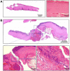
Figure 3. Pathological findings of the resected appendix. (A) Hematoxylin and eosin staining of the fish bone. The fish bone comprises eosinophilic lamellae of mature bone without viable osteocytes. (B) Hematoxylin and eosin staining of the appendix. A transmural cut line of approximately 0.5 mm that is surrounded by fibrous tissue with inflammation is seen.

References

    1. Maleki M, Evans WE. Foreign-body perforation of the intestinal tract. Report of 12 cases and review of the literature. Arch Surg. 1970;101(4):475–477. doi: 10.1001/archsurg.1970.01340280027008. - DOI - PubMed
    1. Goh BK, Chow PK, Quah HM, Ong HS, Eu KW, Ooi LL, Wong WK. Perforation of the gastrointestinal tract secondary to ingestion of foreign bodies. World J Surg. 2006;30(3):372–377. doi: 10.1007/s00268-005-0490-2. - DOI - PubMed
    1. Bunker PG. The role of dentistry in problems of foreign bodies in the air and food passages. J Am Dent Assoc. 1962;64:782–787. doi: 10.14219/jada.archive.1962.0160. - DOI - PubMed
    1. Bhangu A, Søreide K, Di Saverio S, Assarsson JH, Drake FT. Acute appendicitis: modern understanding of pathogenesis, diagnosis, and management. Lancet. 2015;386(10000):1278–1287. doi: 10.1016/S0140-6736(15)00275-5. - DOI - PubMed
    1. Savrin RA, Clausen K, Martin EW Jr, Cooperman M. Chronic and recurrent appendicitis. Am J Surg. 1979;137(3):355–357. doi: 10.1016/0002-9610(79)90066-7. - DOI - PubMed

Publication types