. 2023 Jul;64(7):454-458.
doi: 10.11622/smedj.2022083.

Pembrolizumab-related pneumonitis in a patient with COVID-19 infection

Affiliations

Pembrolizumab-related pneumonitis in a patient with COVID-19 infection

Venkataganesan Ponnalagu et al. Singapore Med J. 2023 Jul.
No abstract available


Conflict of interest statement

None

Figures
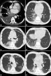
Figure 1
Figure 1
Serial axial CT images of the patient’s thorax (a) before treatment in September 2019; (b & c) after radiotherapy and the initial stage of immunotherapy in March 2020; and in (d) April and (e & f) May 2020. (a) CT image shows a soft tissue mass in the right lower lobe, with an associated right pleural effusion (asterisk). There is invasion of the right lower pulmonary vein (white arrow), right lower lobe segmental bronchus (dashed arrow) and oesophagus by the mass. (b) CT image shows significant reduction in the mass after treatment. Interval development of fibrotic changes is seen along with new ground-glass opacities (GGOs) (dashed arrow). (c) Serial axial CT image of the thorax in March 2020 shows GGOs in the left upper lobe (white arrow). (d) CT image of the thorax shows GGOs and interlobular septal thickening in the right lower lobe (white arrows). (e) CT image shows progression of GGOs in the upper lobes (white arrows) and (f) the left lower lobe posterior segment as well as the left inferior lingular segment (dashed arrows), in keeping with pembrolizumab-related pneumonitis.
Figure 2
Figure 2
Timeline shows the chronology of the imaging scans and COVID-19 swab results of the patient from diagnosis of cancer to the patient’s death. CT TAP: computed tomography thorax, abdomen and pelvis, GGOs: ground-glass opacities, PET-CT: positron emission tomography and CT

References

    1. Wong HYF, Lam HYS, Fong AHT, Leung ST, Chin TW, Lo CSY, et al. Frequency and distribution of chest radiographic findings in patients positive for COVID-19. Radiology. 2020;296:E72–8. - PMC - PubMed
    1. Chung M, Bernheim A, Mei X, Zhang N, Huang M, Zeng X, et al. CT imaging features of 2019 novel coronavirus (2019-nCoV) Radiology. 2020;295:202–7. - PMC - PubMed
    1. Zhou S, Wang Y, Zhu T, Xia L. CT features of coronavirus disease 2019 (COVID-19) pneumonia in 62 patients in Wuhan, China. AJR Am J Roentgenol. 2020;214:1287–94. - PubMed
    1. Bernheim A, Mei X, Huang M, Yang Y, Fayad ZA, Zhang N, et al. Chest CT findings in coronavirus disease-19 (COVID-19): Relationship to duration of infection. Radiology. 2020;295:200463. - PMC - PubMed
    1. Kanne JP, Little BP, Chung JH, Elicker BM, Ketai LH. Essentials for radiologists on COVID-19: An update—Radiology Scientific Expert Panel. Radiology. 2020;296:E113–4. - PMC - PubMed

Substances